Explore
 Validate
 Learn
LearnPA5-23062
antibody from Invitrogen Antibodies
Targeting: CX3CL1
ABCD-3, C3Xkine, CXC3, CXC3C, NTN, SCYD1
 Western blot
Antibody data
- Antibody Data
- Antigen structure
- References [3]
- Comments [0]
- Validations
- Western blot [1]
- Immunocytochemistry [1]
- Immunohistochemistry [2]
- Other assay [3]
Submit
Validation data
Reference
Comment
Report error
- Product number
- PA5-23062 - Provider product page

- Provider
- Invitrogen Antibodies
- Product name
- CX3CL1 Polyclonal Antibody
- Antibody type
- Polyclonal
- Antigen
- Synthetic peptide
- Reactivity
- Human, Mouse
- Host
- Rabbit
- Isotype
- IgG
- Vial size
- 100 µL
- Concentration
- 1.15 mg/mL
- Storage
- Store at 4°C short term. For long term storage, store at -20°C, avoiding freeze/thaw cycles.
Submitted references Insights Into the Somatic Mutation Burden of Hepatoblastomas From Brazilian Patients.
CX(3)CR1-CX(3)CL1-dependent cell-to-cell Japanese encephalitis virus transmission by human microglial cells.
Augmented concentrations of CX3CL1 are associated with interstitial lung disease in systemic sclerosis.
Aguiar TFM, Rivas MP, Costa S, Maschietto M, Rodrigues T, Sobral de Barros J, Barbosa AC, Valieris R, Fernandes GR, Bertola DR, Cypriano M, Caminada de Toledo SR, Major A, Tojal I, Apezzato MLP, Carraro DM, Rosenberg C, Lima da Costa CM, Cunha IW, Sarabia SF, Terrada DL, Krepischi ACV
Frontiers in oncology 2020;10:556
Frontiers in oncology 2020;10:556
CX(3)CR1-CX(3)CL1-dependent cell-to-cell Japanese encephalitis virus transmission by human microglial cells.
Lannes N, Garcia-Nicolàs O, Démoulins T, Summerfield A, Filgueira L
Scientific reports 2019 Mar 18;9(1):4833
Scientific reports 2019 Mar 18;9(1):4833
Augmented concentrations of CX3CL1 are associated with interstitial lung disease in systemic sclerosis.
Hoffmann-Vold AM, Weigt SS, Palchevskiy V, Volkmann E, Saggar R, Li N, Midtvedt Ø, Lund MB, Garen T, Fishbein MC, Ardehali A, Ross DJ, Ueland T, Aukrust P, Lynch JP 3rd, Elashoff RM, Molberg Ø, Belperio JA
PloS one 2018;13(11):e0206545
PloS one 2018;13(11):e0206545
No comments: Submit comment
Supportive validation
- Submitted by
- Invitrogen Antibodies (provider)
- Main image
- Experimental details
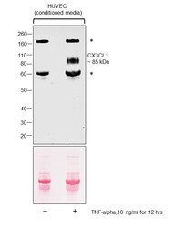
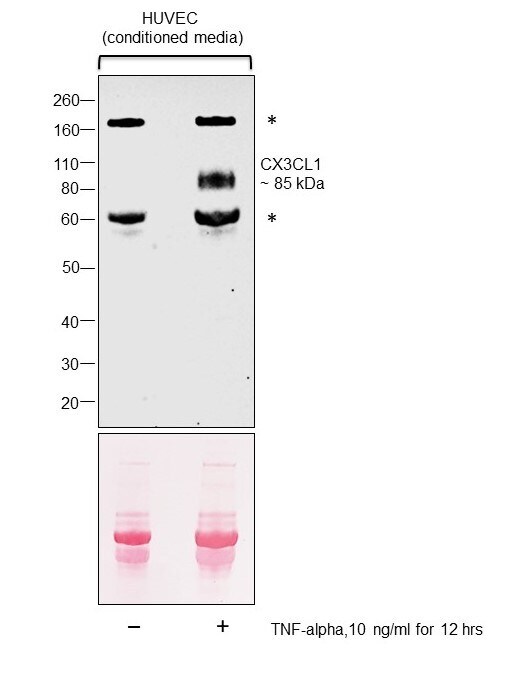
- Western blot was performed using Anti-CX3CL1 Polyclonal Antibody (Product # PA5-23062) and a 85 kDa band corresponding to soluble cleaved form of CX3CL1 was observed across tested cell line along with uncharacterized (*) bands at ~180 kDa and 60 kDa. Conditioned media (20 µg lysate) of HUVEC (Lane 1), HUVEC treated with TNF-alpha, 10 ng/ml for 12 hrs (Lane 2) were electrophoresed using NuPAGE™ 4-12% Bis-Tris Protein Gel (Product # NP0321BOX). Resolved proteins were then transferred onto a Nitrocellulose membrane (Product # IB23001) by iBlot® 2 Dry Blotting System (Product # IB21001). The blot was probed with the primary antibody (1:1000 dilution) and detected by chemiluminescence with Goat anti-Rabbit IgG (H+L) Superclonal™ Recombinant Secondary Antibody, HRP (Product # A27036, 1:4000 dilution) using the iBright FL 1000 (Product # A32752). Chemiluminescent detection was performed using Novex® ECL Chemiluminescent Substrate Reagent Kit (Product # WP20005).
Supportive validation
- Submitted by
- Invitrogen Antibodies (provider)
- Main image
- Experimental details
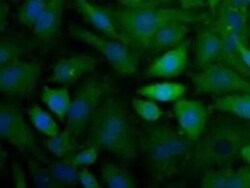
- Immunocytochemistry analysis of CX3CL1 in HeLa cells. Samples were incubated in CX3CL1 polyclonal antibody (Product # PA5-23062). CX3CL (green). Nuclei (Blue) are counterstained using Hoechst 33258..

Supportive validation
- Submitted by
- Invitrogen Antibodies (provider)
- Main image
- Experimental details
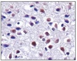
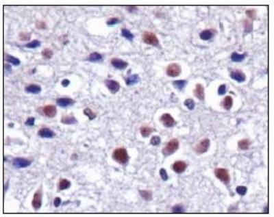
- Immunohistochemical analysis of CX3CL1 in mouse brain. Samples were incubated in CX3CL1 polyclonal antibody (Product # PA5-23062).
- Submitted by
- Invitrogen Antibodies (provider)
- Main image
- Experimental details
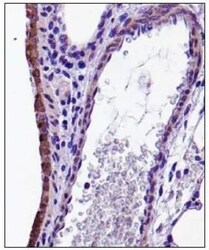
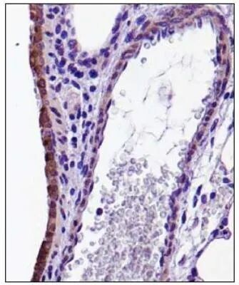
- Immunohistochemical analysis of CX3CL1 in mouse lung. Samples were incubated in CX3CL1 polyclonal antibody (Product # PA5-23062).
Supportive validation
- Submitted by
- Invitrogen Antibodies (provider)
- Main image
- Experimental details
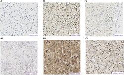
- Figure 3 Protein expression of CX3CL1 and CX3CR1 evaluated in hepatoblastoma samples by immunohistochemistry assay. (A-C) Show CX3CR1 data, and (A1-C1) , CX3CL1 from the same tumor samples. (A) HB17, example of negative labeling for CX3CR1 (A) and CX3CL1 (A1) . (B) HB32T, positive for nuclear and cytoplasmic CX3CR1 (B) and CX3CL1 (B1) . (C) HB33T, positive for cytoplasmic CX3CR1 labeling (C) and positive for nuclear and cytoplasmic CX3CL1 (C1) .
- Submitted by
- Invitrogen Antibodies (provider)
- Main image
- Experimental details
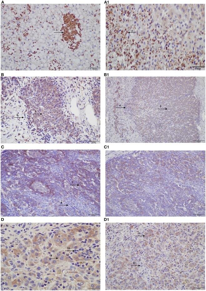
- Figure 4 Protein expression of CX3CL1 and CX3CR1 evaluated in hepatoblastomas and hepatoblastoma lung metastasis by immunohistochemistry assay. (A-D) Show CX3CL1 data, and (A1-D1) , CX3CR1. (A) TCH361, CX3CL1 strong positivity of infiltrated lymphocytes (indicated by arrow 1) in necrotic regions of the tumor, and (A1) , CX3CR1 strong positivity of infiltrated lymphocytes (indicated by arrow 2) in necrotic regions of the tumor; (B,B1) TCH327, positivity in tumor cells (indicated by arrows 3 and 5) and infiltrated lymphocytes negative (indicated by arrows 4 and 6) for both proteins. (C) TCH321, positivity in the osteoblast component and strong positivity in the fetal type (indicated by arrow 7); infiltrated lymphocytes are negative (indicated by arrow 8); (C1) positivity in tumor cells and lymphocytes negative; (D,D1) TCH360, lung metastasis showing positivity in tumor cells (indicated by arrows 9 and 11), and no expression in infiltrated lymphocytes (indicated by arrows 10 and 12), for both proteins.
- Submitted by
- Invitrogen Antibodies (provider)
- Main image
- Experimental details
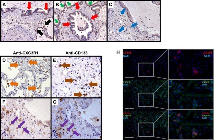
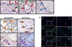
- Fig 2 Immunostaining of CX 3 CL1 and CX 3 CR1 in SSc-ILD. CX 3 CL1 protein is produced from the epithelium and infiltrating interstitial leukocytes in SSc-ILD, while its receptor CX 3 CR1 is localized to infiltrating mononuclear cells. Representative histopathological staining of (n = 5) SSc-ILD for CX 3 CL1 from (A) Type 2 pneumocytes, (B) Airway epithelium, (C) Epithelium involved in bronchiolization, and (D) Infiltrating mononuclear cells. Representative histopathological staining of (n = 5) SSc-ILD for CX 3 CR1 from (E) Infiltrating interstitial mononuclear cells, (F) Morphologically from plasma cells via their eccentric cartwheel nuclei, (G) Representative staining of plasma cell marker CD138 confirming that the cells with eccentric cartwheel nuclei are plasma cells, (H) Representative immunofluorescence staining of plasma cell (red) in the interstitium, CX 3 CR1 positive infiltrating mononuclear cells, and co-localization of CD138 + plasma cell expressing CX 3 CR1 and infiltrating the interstitium.