Antibody data
- Antibody Data
- Antigen structure
- References [0]
- Comments [0]
- Validations
- Western blot [1]
- Immunohistochemistry [12]
Submit
Validation data
Reference
Comment
Report error
- Product number
- HPA021011 - Provider product page
- Provider
- Atlas Antibodies
- Proper citation
- Atlas Antibodies Cat#HPA021011, RRID:AB_1848532
- Product name
- Anti-FGL2
- Antibody type
- Polyclonal
- Reactivity
- Human
- Host
- Rabbit
- Conjugate
- Unconjugated
- Antigen sequence
RNGLLLPSTGAPGEVGDNRVRELESEVNKLSSELK
NAKEEINVLHGRLEKLNLVNMNNIENYVDSKVANL
TFVVNSLDGKCSKCP- Isotype
- IgG
- Vial size
- 100 µl
- Storage
- Store at +4°C for short term storage. Long time storage is recommended at -20°C.
No comments: Submit comment
Supportive validation
- Submitted by
- Atlas Antibodies (provider)
- Main image
- Experimental details
- Lane 1: Marker [kDa] 230, 130, 95, 72, 56, 36, 28, 17, 11Lane 2: Human cell line RT-4
Enhanced validation
Enhanced validation
Supportive validation
- Submitted by
- Atlas Antibodies (provider)
- Enhanced method
- Orthogonal validation
- Main image
- Experimental details
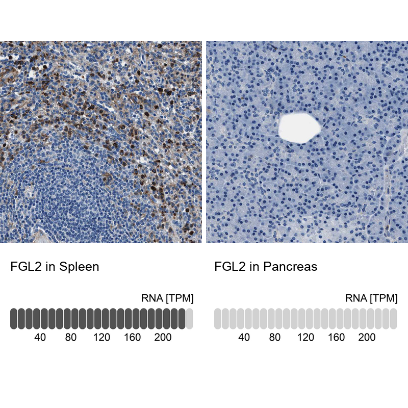
- Immunohistochemistry analysis in human spleen and pancreas tissues using HPA021011 antibody. Corresponding FGL2 RNA-seq data are presented for the same tissues.
- Sample type
- HUMAN
Enhanced validation
- Submitted by
- Atlas Antibodies (provider)
- Enhanced method
- Independent antibody validation
- Main image
- Experimental details
- Immunohistochemical staining of human cerebellum, lymph node, skeletal muscle and spleen using Anti-FGL2 antibody HPA021011 (A) shows similar protein distribution across tissues to independent antibody HPA026682 (B).
Supportive validation
- Submitted by
- Atlas Antibodies (provider)
- Main image
- Experimental details
- Immunohistochemical staining of human spleen shows strong cytoplasmic positivity in cells of red pulp.
- Submitted by
- Atlas Antibodies (provider)
- Main image
- Experimental details
- Immunohistochemical staining of human spleen shows high expression.
- Sample type
- HUMAN
- Submitted by
- Atlas Antibodies (provider)
- Main image
- Experimental details
- Immunohistochemical staining of human skeletal muscle shows low expression as expected.
- Sample type
- HUMAN
- Submitted by
- Atlas Antibodies (provider)
- Main image
- Experimental details
- Immunohistochemical staining of human liver using Anti-FGL2 antibody HPA021011.
- Sample type
- HUMAN
- Submitted by
- Atlas Antibodies (provider)
- Main image
- Experimental details
- Immunohistochemical staining of human lymph node using Anti-FGL2 antibody HPA021011.
- Sample type
- HUMAN
- Submitted by
- Atlas Antibodies (provider)
- Main image
- Experimental details
- Immunohistochemical staining of human spleen shows strong cytoplasmic positivity in cells in red pulp.
- Sample type
- HUMAN
- Submitted by
- Atlas Antibodies (provider)
- Main image
- Experimental details
- Immunohistochemical staining of human pancreas shows no positivity in exocrine glandular cells as expected.
- Sample type
- HUMAN
- Submitted by
- Atlas Antibodies (provider)
- Main image
- Experimental details
- Immunohistochemical staining of human lymph node shows strong cytoplasmic positivity in non-germinal center cells.
- Sample type
- HUMAN
- Submitted by
- Atlas Antibodies (provider)
- Main image
- Experimental details
- Immunohistochemical staining of human skeletal muscle shows no positivity in myocytes as expected.
- Sample type
- HUMAN
- Submitted by
- Atlas Antibodies (provider)
- Main image
- Experimental details
- Immunohistochemical staining of human cerebellum shows moderate cytoplasmic positivity in Purkinje cells.
- Sample type
- HUMAN