Explore
 Validate
 Learn
Learn Western blot
 Immunocytochemistry
Antibody data
- Antibody Data
- Antigen structure
- References [6]
- Comments [0]
- Validations
- Immunocytochemistry [1]
Submit
Validation data
Reference
Comment
Report error
- Product number
- HPA036565 - Provider product page

- Provider
- Atlas Antibodies
- Proper citation
- Atlas Antibodies Cat#HPA036565, RRID:AB_10673623
- Product name
- Anti-MATR3
- Antibody type
- Polyclonal
- Description
- Polyclonal Antibody against Human MATR3, Gene description: matrin 3, Alternative Gene Names: KIAA0723, MGC9105, MPD2, VCPDM, Validated applications: WB, IHC, ICC, Uniprot ID: P43243, Storage: Store at +4°C for short term storage. Long time storage is recommended at -20°C.
- Reactivity
- Human, Mouse
- Host
- Rabbit
- Conjugate
- Unconjugated
- Isotype
- IgG
- Vial size
- 100 µl
- Concentration
- 0.1 mg/ml
- Storage
- Store at +4°C for short term storage. Long time storage is recommended at -20°C.
- Handling
- The antibody solution should be gently mixed before use.
Submitted references MATR3 P154S knock-in mice do not exhibit motor, muscle or neuropathologic features of ALS
Low Expression of MATR3 Is Associated with Poor Survival in Clear Cell Renal Cell Carcinoma
Revealing protein-protein interactions at the transcriptome scale by sequencing.
Selective neuronal degeneration in MATR3 S85C knock-in mouse model of early-stage ALS
ALS Associated Mutations in Matrin 3 Alter Protein-Protein Interactions and Impede mRNA Nuclear Export
Mutations in the Matrin 3 gene cause familial amyotrophic lateral sclerosis
Dominick M, Houchins N, Venugopal V, Zuberi A, Lutz C, Meechooveet B, Van Keuren-Jensen K, Bowser R, Medina D
Biochemical and Biophysical Research Communications 2023;645
Biochemical and Biophysical Research Communications 2023;645
Low Expression of MATR3 Is Associated with Poor Survival in Clear Cell Renal Cell Carcinoma
Durślewicz J, Klimaszewska-Wiśniewska A, Antosik P, Grzanka D
Biomedicines 2023;11(2):326
Biomedicines 2023;11(2):326
Revealing protein-protein interactions at the transcriptome scale by sequencing.
Johnson KL, Qi Z, Yan Z, Wen X, Nguyen TC, Zaleta-Rivera K, Chen CJ, Fan X, Sriram K, Wan X, Chen ZB, Zhong S
Molecular cell 2021 Oct 7;81(19):4091-4103.e9
Molecular cell 2021 Oct 7;81(19):4091-4103.e9
Selective neuronal degeneration in MATR3 S85C knock-in mouse model of early-stage ALS
Kao C, van Bruggen R, Kim J, Chen X, Chan C, Lee J, Cho W, Zhao M, Arndt C, Maksimovic K, Khan M, Tan Q, Wilson M, Park J
Nature Communications 2020;11(1)
Nature Communications 2020;11(1)
ALS Associated Mutations in Matrin 3 Alter Protein-Protein Interactions and Impede mRNA Nuclear Export
Boehringer A, Garcia-Mansfield K, Singh G, Bakkar N, Pirrotte P, Bowser R
Scientific Reports 2017;7(1)
Scientific Reports 2017;7(1)
Mutations in the Matrin 3 gene cause familial amyotrophic lateral sclerosis
Johnson J, Pioro E, Boehringer A, Chia R, Feit H, Renton A, Pliner H, Abramzon Y, Marangi G, Winborn B, Gibbs J, Nalls M, Morgan S, Shoai M, Hardy J, Pittman A, Orrell R, Malaspina A, Sidle K, Fratta P, Harms M, Baloh R, Pestronk A, Weihl C, Rogaeva E, Zinman L, Drory V, Borghero G, Mora G, Calvo A, Rothstein J, Drepper C, Sendtner M, Singleton A, Taylor J, Cookson M, Restagno G, Sabatelli M, Bowser R, Chiò A, Traynor B
Nature Neuroscience 2014;17(5):664-666
Nature Neuroscience 2014;17(5):664-666
No comments: Submit comment
Supportive validation
- Submitted by
- Atlas Antibodies (provider)
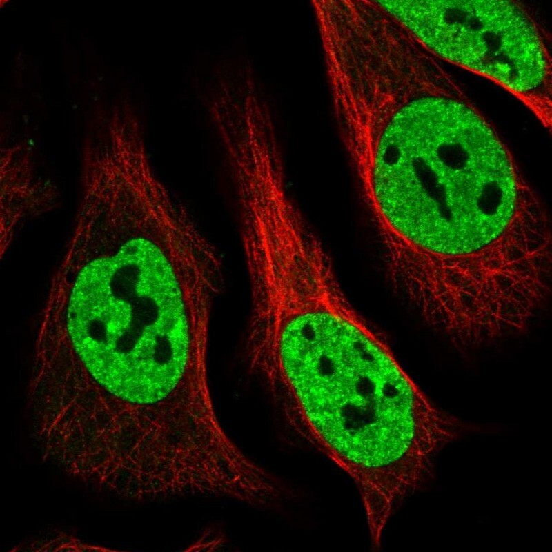
- Main image
- Experimental details
- Immunofluorescent staining of human cell line U-2 OS shows localization to nucleoplasm.
- Sample type
- Human