 Explore
 Validate
 Learn
Learn Western blot
 Immunocytochemistry
Antibody data
- Antibody Data
- Antigen structure
- References [0]
- Comments [0]
- Validations
- Immunocytochemistry [4]
- Immunoprecipitation [1]
- Immunohistochemistry [4]
- Other assay [1]
Submit
Validation data
Reference
Comment
Report error
- Product number
- PA5-57720 - Provider product page

- Provider
- Invitrogen Antibodies
- Product name
- MATR3 Polyclonal Antibody
- Antibody type
- Polyclonal
- Antigen
- Recombinant protein fragment
- Description
- Immunogen sequence: LKRRRTEEGP TLSYGRDGRS ATREPPYRVP RDDWEEKRHF RRDSFDDRGP SLNPVLDYDH GSRSQESGYY DRMDYEDDRL RDGERCRDD Highest antigen sequence identity to the following orthologs: Mouse - 100%, Rat - 100%.
- Reactivity
- Human, Mouse
- Host
- Rabbit
- Isotype
- IgG
- Vial size
- 100 μL
- Concentration
- 0.1 mg/mL
- Storage
- Store at 4°C short term. For long term storage, store at -20°C, avoiding freeze/thaw cycles.
No comments: Submit comment
Supportive validation
- Submitted by
- Invitrogen Antibodies (provider)
- Main image
- Experimental details
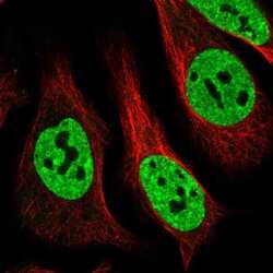
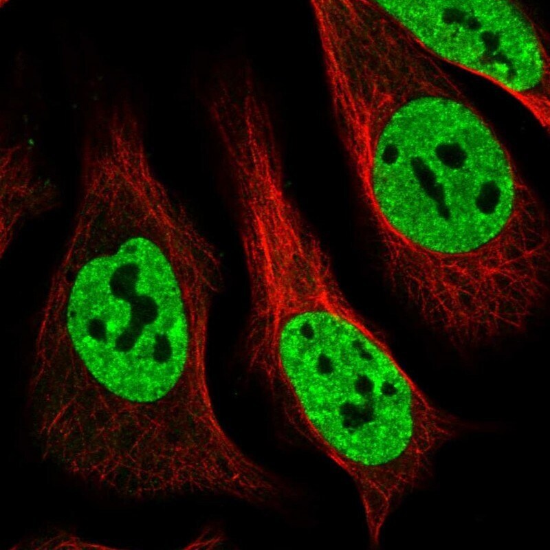
- Immunofluorescent staining of MATR3 in human cell line U-2 OS shows positivity in nucleus but excluded from the nucleoli. Samples were probed using a MATR3 Polyclonal Antibody (Product # PA5-57720).

- Submitted by
- Invitrogen Antibodies (provider)
- Main image
- Experimental details
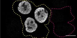
- Immunofluorescence of MATR3 was performed using HAP1 wild-type and MATR3 KO cells that were transfected with a green or a far-red fluorescent dye, respectively. Post-transfection, WT and KO cells were mixed and plated to a 1:1 ratio on coverslips as a mosaic and incubated for 24 hrs. Cells were fixed in 4% PFA (in PBS) for 15 min; cells were permeabilized with 0.1% Triton X-100 for 10 min at RT and blocked with PBS with 5% BSA, 5% goat serum, and 0.01% Triton X-100 for 30 min. Cells were stained with the MATR3 polyclonal antibody (Product # PA5-57720) at a 1:2,000 dilution overnight at 4°C. Secondary antibody incubation was performed using 1 µg/mL of Goat anti-Rabbit IgG (H+L) Highly Cross-Adsorbed Secondary Antibody, Alexa Fluor 555 antibody (Product # A21429) for 1 hr. Imaging was performed with a 40X oil objective and analysis was performed using Image J. Cell image represents a single focal plane; WT and KO cells are outlined with a yellow (WT) or magenta (KO) dashed line. Data courtesy of YCharOS Inc., an open science company with the mission of characterizing commercially available antibodies using knockout validation.

- Submitted by
- Invitrogen Antibodies (provider)
- Main image
- Experimental details
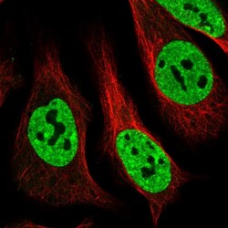
- Immunofluorecent analysis of MATR3 in human cell line U-2 OS using MATR3 Polyclonal Antibody (Product # PA5-57720). Staining shows localization to nucleoplasm.
- Submitted by
- Invitrogen Antibodies (provider)
- Main image

- Experimental details
- Immunofluorescence of MATR3 was performed using HAP1 wild-type and MATR3 KO cells that were transfected with a green or a far-red fluorescent dye, respectively. Post-transfection, WT and KO cells were mixed and plated to a 1:1 ratio on coverslips as a mosaic and incubated for 24 hrs. Cells were fixed in 4% PFA (in PBS) for 15 min; cells were permeabilized with 0.1% Triton X-100 for 10 min at RT and blocked with PBS with 5% BSA, 5% goat serum, and 0.01% Triton X-100 for 30 min. Cells were stained with the MATR3 polyclonal antibody (Product # PA5-57720) at a 1:2,000 dilution overnight at 4°C. Secondary antibody incubation was performed using 1 µg/mL of Goat anti-Rabbit IgG (H+L) Highly Cross-Adsorbed Secondary Antibody, Alexa Fluor 555 antibody (Product # A21429) for 1 hr. Imaging was performed with a 40X oil objective and analysis was performed using Image J. Cell image represents a single focal plane; WT and KO cells are outlined with a yellow (WT) or magenta (KO) dashed line. Data courtesy of YCharOS Inc., an open science company with the mission of characterizing commercially available antibodies using knockout validation.

Supportive validation
- Submitted by
- Invitrogen Antibodies (provider)
- Main image
- Experimental details
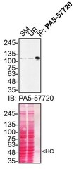
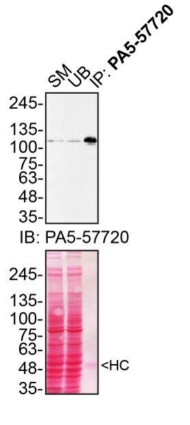
- Immunoprecipitation of MATR3 was performed on HAP1 cell lysates. Antibody-bead conjugates were prepared by adding 2 µg of MATR3 polyclonal antibody (Product # PA5-57720) with 30 µL of protein A-Sepharose beads and rocked overnight at 4°C. 1 mg of lysate was incubated with an antibody-bead conjugate for 2 hours at 4°C. Following centrifugation and multiple washes, 10% starting material (SM), 10% unbound fraction (UB) and immunoprecipitated fraction (IP) were processed for immunoblot using the same antibody. Ponceau stained transfer of blot is shown. Data courtesy of YCharOS Inc., an open science company with the mission of characterizing commercially available antibodies using knockout validation.
Supportive validation
- Submitted by
- Invitrogen Antibodies (provider)
- Main image

- Experimental details
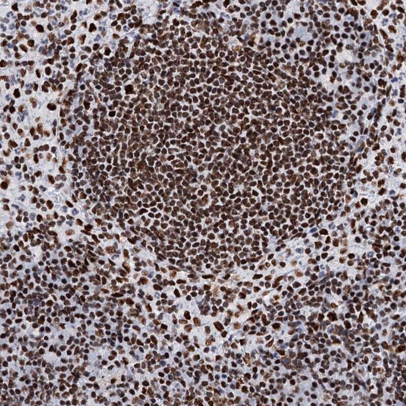
- Immunohistochemical staining of MATR3 in human lymph node using MATR3 Polyclonal Antibody (Product # PA5-57720) shows moderate to strong nuclear positivity in lymphoid cells.
- Submitted by
- Invitrogen Antibodies (provider)
- Main image
- Experimental details
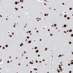
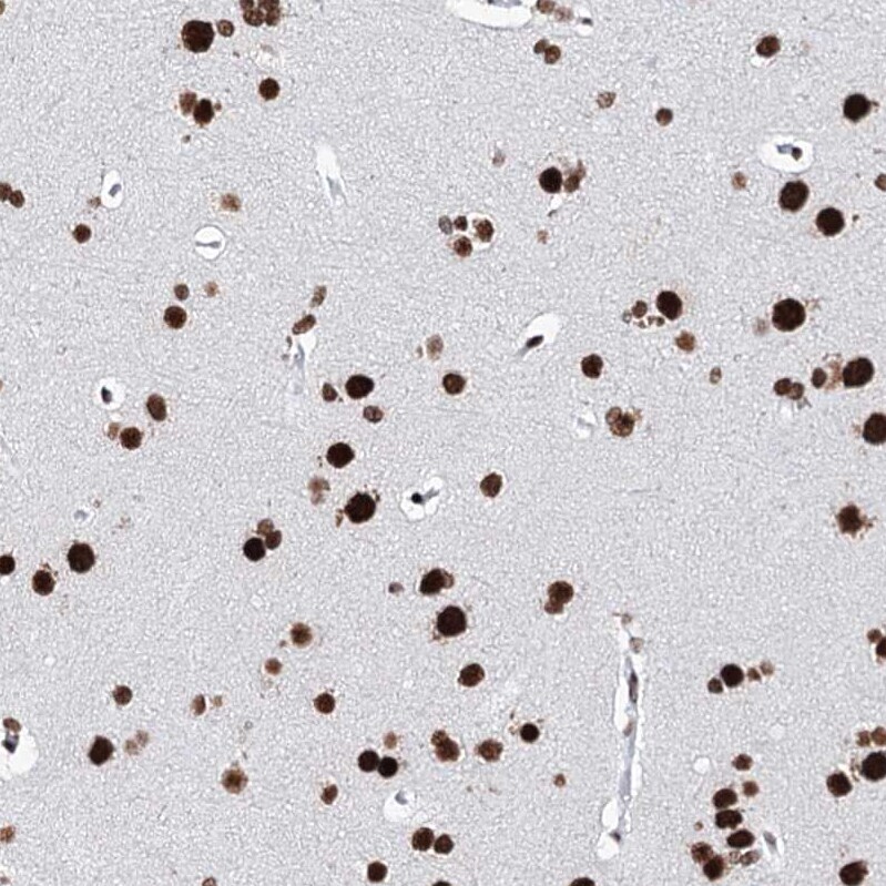
- Immunohistochemical staining of MATR3 in human cerebral cortex using MATR3 Polyclonal Antibody (Product # PA5-57720) shows strong nuclear positivity in neurons.
- Submitted by
- Invitrogen Antibodies (provider)
- Main image
- Experimental details
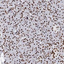
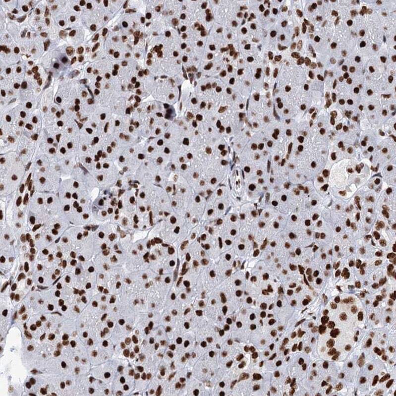
- Immunohistochemical staining of MATR3 in human pancreas using MATR3 Polyclonal Antibody (Product # PA5-57720) shows moderate nuclear positivity in exocrine glandular cells.
- Submitted by
- Invitrogen Antibodies (provider)
- Main image
- Experimental details
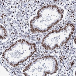
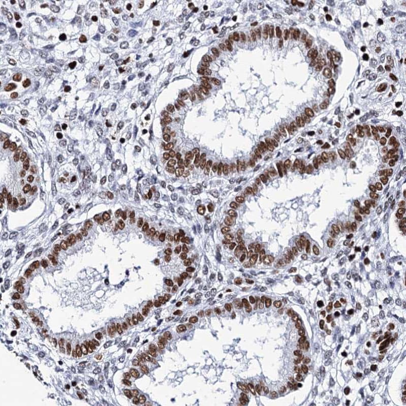
- Immunohistochemical staining of MATR3 in human endometrium using MATR3 Polyclonal Antibody (Product # PA5-57720) shows moderate nuclear positivity in glandular cells.
Supportive validation
- Submitted by
- Invitrogen Antibodies (provider)
- Main image

- Experimental details
- Immunoprecipitation of MATR3 was performed on HAP1 cell lysates. Antibody-bead conjugates were prepared by adding 2 µg of MATR3 polyclonal antibody (Product # PA5-57720) with 30 µL of protein A-Sepharose beads and rocked overnight at 4°C. 1 mg of lysate was incubated with an antibody-bead conjugate for 2 hours at 4°C. Following centrifugation and multiple washes, 10% starting material (SM), 10% unbound fraction (UB) and immunoprecipitated fraction (IP) were processed for immunoblot using the same antibody. Ponceau stained transfer of blot is shown. Data courtesy of YCharOS Inc., an open science company with the mission of characterizing commercially available antibodies using knockout validation.
