Explore
 Validate
 Learn
Learn Flow cytometry
Antibody data
- Antibody Data
- Antigen structure
- References [0]
- Comments [0]
- Validations
- Flow cytometry [2]
Submit
Validation data
Reference
Comment
Report error
- Product number
- 17-9038-42 - Provider product page

- Provider
- Invitrogen Antibodies
- Product name
- Phospho-MCL-1 (Ser159) Monoclonal Antibody (RBCERNR), APC, eBioscience™
- Antibody type
- Monoclonal
- Antigen
- Other
- Description
- Description: This RBCERNR monoclonal antibody recognizes human and mouse myeloid cell leukemia sequence 1 (Mcl-1) when phosphorylated on serine 159 (S159). Mcl-1 is an anti-apoptotic protein that is a member of the Bcl-2 family of proteins important for regulation of cell survival/apoptosis. Mcl-1 is primarily localized to the outer membrane of mitochondria where it prevents cytochrome c release via dimerization with other Bcl-2 family members such as Bim. PI3K activation of AKT results in the phosphorylation of GSK3 beta at serine 9 (S9) resulting in destabilization and degradation of GSK3 beta. Loss of GSK3 beta prevents phosphorylation of Mcl-1 on S159 and its subsequent ubiquitination and degradation. Mice conditionally lacking Mcl-1 in lymphocytes showed that Mcl-1 is essential during early lymphoid development and for the maintenance of mature lymphocytes. Applications Reported:This RBCERNR antibody has been reported for use in intracellular staining followed by flow cytometric analysis. Applications Tested: This RBCERNR antibody has been pre-titrated and tested by intracellular staining followed by flow cytometric analysis of normal human peripheral blood cells. This can be used at 5 µL (0.25 µg) per test. A test is defined as the amount (µg) of antibody that will stain a cell sample in a final volume of 100 µL. Cell number should be determined empirically but can range from 10^5 to 10^8 cells/test. Staining Protocol: Protocol A and Protocol C are recommended for this monoclonal antibody. Use of Protocol A: Two-step protocol: intracellular (cytoplasmic) proteins allows for the greatest flexibility for detection of surface and intracellular (cytoplasmic) proteins. Protocol C: Two-step protocol: Fixation/Methanol allows for the greatest discrimination of phospho-specific signaling between unstimulated and stimulated samples, but with limitations on the ability to stain specific surface proteins (refer to "Clone Performance Following Fixation/Permeabilization" located in the BestProtocols Section under the Resources tab online). All Protocols can be found in the Flow Cytometry Protocols: "Staining Intracellular Antigens for Flow Cytometry Protocol" located in the BestProtocols® Section under the Resources tab online. Excitation: 633-647 nm; Emission: 660 nm; Laser: Red Laser. Filtration: 0.2 µm post-manufacturing filtered.
- Reactivity
- Human, Mouse
- Host
- Mouse
- Isotype
- IgG
- Antibody clone number
- RBCERNR
- Vial size
- 100 Tests
- Concentration
- 5 µL/Test
- Storage
- 4°C, store in dark, DO NOT FREEZE!
No comments: Submit comment
Supportive validation
- Submitted by
- Invitrogen Antibodies (provider)
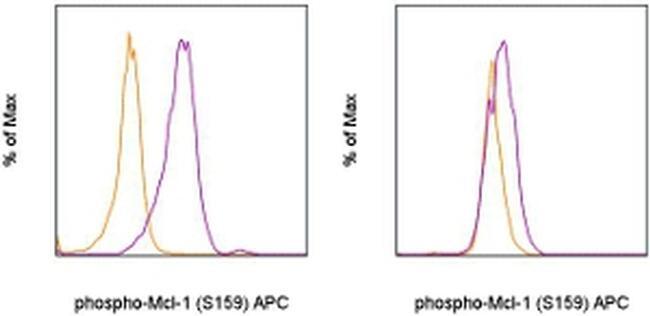
- Main image
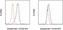
- Experimental details
- Normal human peripheral blood cells were unstimulated (orange histogram) or were stimulated with Anti-Human CD3 and CD28 Functional Grade Purifieds (Product # 16-0037-81 and Product # 16-0289-81) in the presence of the proteasome inhibitor, MG-132 (purple histogram). The cells were then intracellularly stained with Anti-Human/Mouse phospho-Mcl-1 (S159) APC and Anti-Human CD3 PerCP-Cyanine5-5 (Product # 45-0036-42) (left) or Anti-Human CD19 PE (Product # 12-0199-42) (right) using the Intracellular Fixation & Permeabilization Buffer Set (Product # 88-8824-00) and protocol. Cells in the lymphocyte gate were used for analysis.
- Submitted by
- Invitrogen Antibodies (provider)
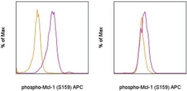
- Main image
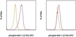
- Experimental details
- Normal human peripheral blood cells were unstimulated (orange histogram) or were stimulated with Anti-Human CD3 and CD28 Functional Grade Purifieds (Product # 16-0037-81 and Product # 16-0289-81) in the presence of the proteasome inhibitor, MG-132 (purple histogram). The cells were then intracellularly stained with Anti-Human/Mouse phospho-Mcl-1 (S159) APC and Anti-Human CD3 PerCP-Cyanine5-5 (Product # 45-0036-42) (left) or Anti-Human CD19 PE (Product # 12-0199-42) (right) using the Intracellular Fixation & Permeabilization Buffer Set (Product # 88-8824-00) and protocol. Cells in the lymphocyte gate were used for analysis.