Explore
 Validate
 Learn
Learn Western blot
 Immunohistochemistry
Antibody data
- Antibody Data
- Antigen structure
- References [1]
- Comments [0]
- Validations
- Western blot [2]
Submit
Validation data
Reference
Comment
Report error
- Product number
- MAB828 - Provider product page

- Provider
- R&D Systems
- Product name
- Human Mcl-1 Antibody
- Antibody type
- Monoclonal
- Description
- Protein A or G purified from hybridoma culture supernatant. Detects human Mcl-1.
- Reactivity
- Human
- Host
- Mouse
- Conjugate
- Unconjugated
- Antigen sequence
Q07820- Isotype
- IgG
- Antibody clone number
- 542808
- Vial size
- 100 ug
- Storage
- Use a manual defrost freezer and avoid repeated freeze-thaw cycles. 12 months from date of receipt, -20 to -70 °C as supplied. 1 month, 2 to 8 °C under sterile conditions after reconstitution. 6 months, -20 to -70 °C under sterile conditions after reconstitution.
Submitted references High-Complexity shRNA Libraries and PI3 Kinase Inhibition in Cancer: High-Fidelity Synthetic Lethality Predictions.
Mues M, Karra L, Romero-Moya D, Wandler A, Hangauer MJ, Ksionda O, Thus Y, Lindenbergh M, Shannon K, McManus MT, Roose JP
Cell reports 2019 Apr 9;27(2):631-647.e5
Cell reports 2019 Apr 9;27(2):631-647.e5
No comments: Submit comment
Supportive validation
- Submitted by
- R&D Systems (provider)
- Main image
- Experimental details
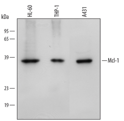
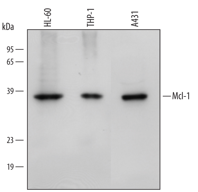
- Detection of Human Mcl-1 by Western Blot. Western blot shows lysates of HL-60 human acute promyelocytic leukemia cell line, THP-1 human acute monocytic leukemia cell line, and A431 human epithelial carcinoma cell line. PVDF membrane was probed with 1 µg/mL of Mouse Anti-Human Mcl-1 Monoclonal Antibody (Catalog # MAB828) followed by HRP-conjugated Anti-Mouse IgG Secondary Antibody (Catalog # HAF007). A specific band was detected for Mcl-1 at approximately 38 kDa (as indicated). This experiment was conducted under reducing conditions and using Immunoblot Buffer Group 2.
- Submitted by
- R&D Systems (provider)
- Main image
- Experimental details
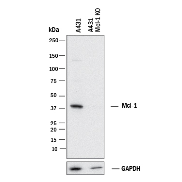
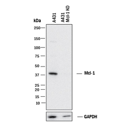
- Western Blot Shows Human Mcl-1 Specificity by Using Knockout Cell Line. Western blot shows lysates of A431 human epithelial carcinoma parental cell line and Mcl-1 knockout A431 cell line (KO). PVDF membrane was probed with 1 µg/mL of Mouse Anti-Human Mcl-1 Monoclonal Antibody (Catalog # MAB828) followed by HRP-conjugated Anti-Mouse IgG Secondary Antibody (Catalog # HAF018). A specific band was detected for Mcl-1 at approximately 40 kDa (as indicated) in the parental A431 cell line, but is not detectable in knockout A431 cell line. GAPDH (Catalog # MAB5718) is shown as a loading control. This experiment was conducted under reducing conditions and using Immunoblot Buffer Group 1.