 Explore
 Validate
 Learn
Learn Western blot
 Other assay
Antibody data
- Antibody Data
- Antigen structure
- References [2]
- Comments [0]
- Validations
- Other assay [2]
Submit
Validation data
Reference
Comment
Report error
- Product number
- PA1-41253 - Provider product page

- Provider
- Invitrogen Antibodies
- Product name
- SOCS5 Polyclonal Antibody
- Antibody type
- Polyclonal
- Antigen
- Synthetic peptide
- Description
- Suggested positive control: human spleen protein, human or mouse spleen, mouse spleen protein.
- Reactivity
- Human, Mouse, Rat
- Host
- Rabbit
- Isotype
- IgG
- Vial size
- 200 μL
- Concentration
- 0.10 mg/mL
- Storage
- Store at 4°C short term. For long term storage, store at -20°C, avoiding freeze/thaw cycles.
Submitted references AMPK suppresses Th2 cell responses by repressing mTORC2.
Angpt2 Induces Mesangial Cell Apoptosis through the MicroRNA-33-5p-SOCS5 Loop in Diabetic Nephropathy.
Pandit M, Timilshina M, Gu Y, Acharya S, Chung Y, Seo SU, Chang JH
Experimental & molecular medicine 2022 Aug;54(8):1214-1224
Experimental & molecular medicine 2022 Aug;54(8):1214-1224
Angpt2 Induces Mesangial Cell Apoptosis through the MicroRNA-33-5p-SOCS5 Loop in Diabetic Nephropathy.
Tsai YC, Kuo PL, Hung WW, Wu LY, Wu PH, Chang WA, Kuo MC, Hsu YL
Molecular therapy. Nucleic acids 2018 Dec 7;13:543-555
Molecular therapy. Nucleic acids 2018 Dec 7;13:543-555
No comments: Submit comment
Supportive validation
- Submitted by
- Invitrogen Antibodies (provider)
- Main image

- Experimental details
- Fig. 6 SOCS5 suppresses GATA3 expression. a Immunoblot analysis of the expression levels of SOCS1, SOCS2, SOCS3, and SOCS5 in wild-type (WT) and AMPK-deficient CD4 + T cells under resting and TCR-stimulated conditions. beta-actin was used as a loading control. The quantified blots are shown as bar graphs in Supplementary Fig. 8a . b , c Immunoblot analysis of SOCS5, p-STAT6, STAT6, GATA3, and the loading control beta-actin with or without TCR stimulation, and IL-4 cytokine, and compound (Comp) C ( b ) or AICAR treatment ( c ). The quantified blots are presented in the bar graphs in Supplementary Fig. 8b , c . d Immunoblot analysis of phosphorylated and total AMPK, SOCS5, GATA3, and the loading control beta-actin in resting and TCR-stimulated WT and Rictor- deficient CD4 + T cells with or without Comp C treatment. The quantified blots are presented in the bar graphs in Supplementary Fig. 8d . e Immunoblot analysis of SOCS5, GATA3, and the loading control beta-actin in resting and TCR-stimulated WT, Sirt1 -deficient, Rictor -deficient, and DKO CD4 + T cells. The quantified blots are represented as bar graphs in Supplementary Fig. 8e . f , g Allergic inflammation was induced in WT, Prkaa1 T-KO ( f ), WT, and Sirt1 T-KO mice with or without AICAR treatment ( g ). Immunoblot analysis of phosphorylated and total STAT6, SOCS5, GATA3, and the loading control beta-actin in CD4 + T cells that were stimulated overnight with OVA. The quantified blots are represented in the bar graphs in

- Submitted by
- Invitrogen Antibodies (provider)
- Main image
- Experimental details
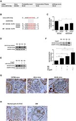
- Figure 5 Suppressor of Cytokine Signaling 5 Is a Direct Target of miR-33-5p in MMCs (A) The predictive binding score of miR-33-5p and suppressor of cytokine signaling 5 (SOCS5) mRNA according to miRmap database. (B) A schematic representation of sequence alignment of SOCS5 mRNA 3' UTR based on TargetScan 7.1 version. (C) Luciferase activity was repressed by endogenous miR-33-5p. HEK293 cells were cotransfected with pGL3-SOCS5-3' UTR luciferase plasmid/pRL-TK Renilla (1:8) or pGL3-SOCS5-3' UTR MT luciferase plasmid/pRL-TK Renilla (1:8) with various miRNA mimics (control mimic or miR-33-5p mimic) by DharmaFECT Duo transfection reagent after 48 hr, and both firefly and Renilla luciferase activities were quantified using the Dual-Glo Luciferase Assay System. Endogenous SOCS5 in MMCs was regulated by miR-33-5p under an HG condition. (D and E) miR-33-5p inhibitor (D) enhanced and miR-33-5p mimic (E) suppressed SOCS5 expression in MMCs under an NG or HG condition. Cells were transfected with either miR-33-5p inhibitor or miR-33-5p mimic, and, 24 hr after transfection, the cells were treated with Angpt2 for 24 hr under an NG or HG condition. Western blotting was utilized to measure SOCS5 protein expression. (F) Angpt2 increased SOCS5 expression in MMCs under an NG condition, and it augmented SOCS5 expression in MMCs under an HG condition. Western blotting and quantitative analysis of SOCS5 was performed in MMCs treated with Angpt2 under NG or HG for 24 hr. (G and H) The expression of
