Explore
 Validate
 Learn
Learn Immunohistochemistry
Antibody data
- Antibody Data
- Antigen structure
- References [5]
- Comments [0]
- Validations
- Immunohistochemistry [1]
Submit
Validation data
Reference
Comment
Report error
- Product number
- HPA008689 - Provider product page

- Provider
- Atlas Antibodies
- Proper citation
- Atlas Antibodies Cat#HPA008689, RRID:AB_1846009
- Product name
- Anti-CPA3
- Antibody type
- Polyclonal
- Description
- Polyclonal Antibody against Human CPA3, Gene description: carboxypeptidase A3 (mast cell), Validated applications: IHC, Uniprot ID: P15088, Storage: Store at +4°C for short term storage. Long time storage is recommended at -20°C.
- Reactivity
- Human
- Host
- Rabbit
- Conjugate
- Unconjugated
- Isotype
- IgG
- Vial size
- 100 µl
- Concentration
- 0.1 mg/ml
- Storage
- Store at +4°C for short term storage. Long time storage is recommended at -20°C.
- Handling
- The antibody solution should be gently mixed before use.
Submitted references Single-cell analysis unveils activation of mast cells in colorectal cancer microenvironment
Carboxypeptidase A3 expression in canine mast cell tumors and tissue-resident mast cells.
Lung Mast Cells Have a High Constitutive Expression of Carboxypeptidase A3 mRNA That Is Independent from Granule-Stored CPA3.
Unique Immune Gene Expression Patterns in Bronchoalveolar Lavage and Tumor Adjacent Non-Neoplastic Lung Tissue in Non-Small Cell Lung Cancer.
Glandular mast cells with distinct phenotype are highly elevated in chronic rhinosinusitis with nasal polyps
Xie Z, Niu L, Zheng G, Du K, Dai S, Li R, Dan H, Duan L, Wu H, Ren G, Dou X, Feng F, Zhang J, Zheng J
Cell & Bioscience 2023;13(1)
Cell & Bioscience 2023;13(1)
Carboxypeptidase A3 expression in canine mast cell tumors and tissue-resident mast cells.
Hämäläinen S, Kareinen L, Sukura A, Kareinen I
Veterinary pathology 2022 Mar;59(2):236-243
Veterinary pathology 2022 Mar;59(2):236-243
Lung Mast Cells Have a High Constitutive Expression of Carboxypeptidase A3 mRNA That Is Independent from Granule-Stored CPA3.
Siddhuraj P, Clausson CM, Sanden C, Alyamani M, Kadivar M, Marsal J, Wallengren J, Bjermer L, Erjefält JS
Cells 2021 Feb 3;10(2)
Cells 2021 Feb 3;10(2)
Unique Immune Gene Expression Patterns in Bronchoalveolar Lavage and Tumor Adjacent Non-Neoplastic Lung Tissue in Non-Small Cell Lung Cancer.
Kuo CS, Liu CY, Pavlidis S, Lo YL, Wang YW, Chen CH, Ko HW, Chung FT, Lin TY, Wang TY, Lee KY, Guo YK, Wang TH, Yang CT
Frontiers in immunology 2018;9:232
Frontiers in immunology 2018;9:232
Glandular mast cells with distinct phenotype are highly elevated in chronic rhinosinusitis with nasal polyps
Takabayashi T, Kato A, Peters A, Suh L, Carter R, Norton J, Grammer L, Tan B, Chandra R, Conley D, Kern R, Fujieda S, Schleimer R
Journal of Allergy and Clinical Immunology 2012;130(2):410-420.e5
Journal of Allergy and Clinical Immunology 2012;130(2):410-420.e5
No comments: Submit comment
Supportive validation
- Submitted by
- Atlas Antibodies (provider)
- Enhanced method
- Orthogonal validation
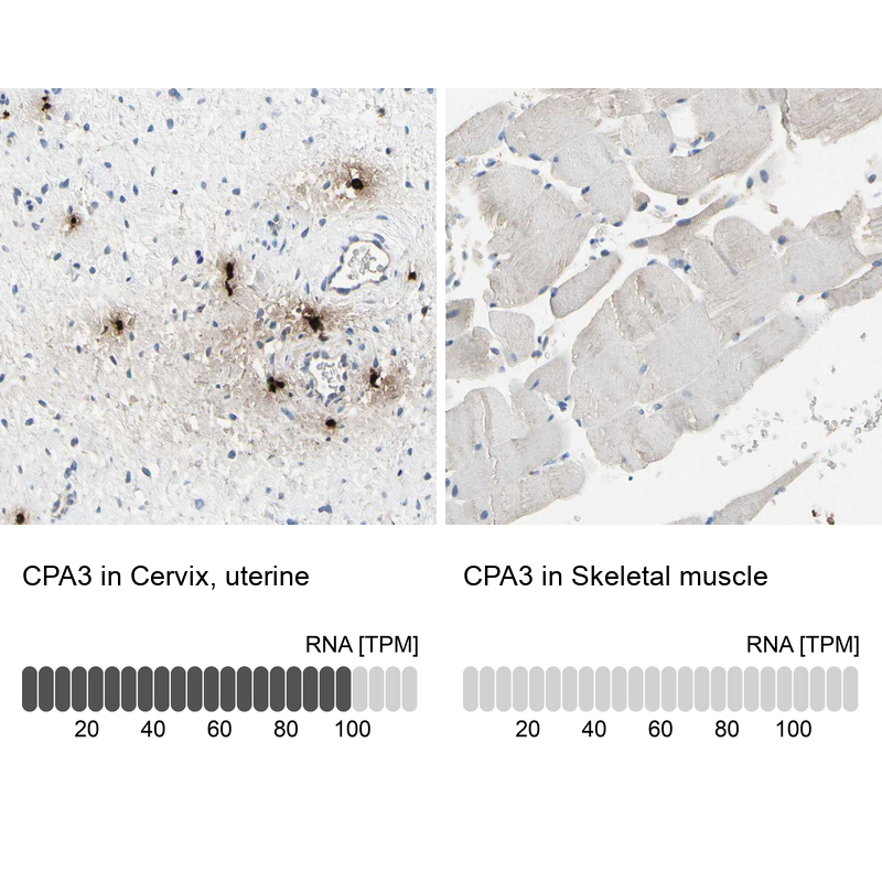
- Main image

- Experimental details
- Immunohistochemistry analysis in human cervix, uterine and skeletal muscle tissues using HPA008689 antibody. Corresponding CPA3 RNA-seq data are presented for the same tissues.
- Sample type
- Human
- Protocol
- Protocol