Explore
 Validate
 Learn
Learn Western blot
 Immunocytochemistry
Antibody data
- Antibody Data
- Antigen structure
- References [0]
- Comments [0]
- Validations
- Immunocytochemistry [3]
Submit
Validation data
Reference
Comment
Report error
- Product number
- GTX82891 - Provider product page

- Provider
- GeneTex
- Proper citation
- GeneTex Cat#GTX82891, RRID:AB_626031
- Product name
- KCNQ2 antibody
- Antibody type
- Polyclonal
- Reactivity
- Human, Mouse, Rat
- Host
- Rabbit
No comments: Submit comment
Supportive validation
- Submitted by
- GeneTex (provider)
- Main image

- Experimental details
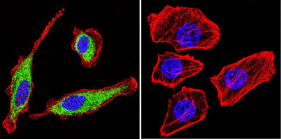
- Immunofluorescent analysis of KCNQ2 using Anti-KCNQ2 Polyclonal Antibody (GTX82891) shows staining in C6 Cells. KCNQ2 staining (green), F-Actin staining with Phalloidin (red) and nuclei with DAPI (blue) is shown. Cells were grown on chamber slides and fixed with formaldehyde prior to staining. Cells were probed without (control) or with or an antibody recognizing KCNQ2 (GTX82891) at a dilution of 1:100 over night at 4 ¢XC, washed with PBS and incubated with a DyLight-488 conjugated secondary antibody (Goat Anti-Rabbit). Images were taken at 60X magnification.
- Submitted by
- GeneTex (provider)
- Main image
- Experimental details
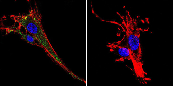
- Immunofluorescent analysis of KCNQ2 using Anti-KCNQ2 Polyclonal Antibody (GTX82891) shows staining in HepG2 Cells. KCNQ2 staining (green), F-Actin staining with Phalloidin (red) and nuclei with DAPI (blue) is shown. Cells were grown on chamber slides and fixed with formaldehyde prior to staining. Cells were probed without (control) or with or an antibody recognizing KCNQ2 (GTX82891) at a dilution of 1:100 over night at 4 ¢XC, washed with PBS and incubated with a DyLight-488 conjugated secondary antibody (Goat Anti-Rabbit). Images were taken at 60X magnification.
- Submitted by
- GeneTex (provider)
- Main image

- Experimental details
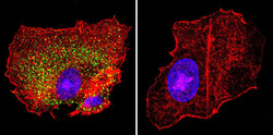
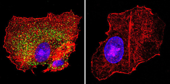
- Immunofluorescent analysis of KCNQ2 using Anti-KCNQ2 Polyclonal Antibody (GTX82891) shows staining in U251 Cells. KCNQ2 staining (green), F-Actin staining with Phalloidin (red) and nuclei with DAPI (blue) is shown. Cells were grown on chamber slides and fixed with formaldehyde prior to staining. Cells were probed without (control) or with or an antibody recognizing KCNQ2 GTX82891 at a dilution of 1:20 over night at 4 ¢XC, washed with PBS and incubated with a DyLight-488 conjugated secondary antibody (Goat Anti-Rabbit). Images were taken at 60X magnification.