Explore
 Validate
 Learn
LearnPA1-929
antibody from Invitrogen Antibodies
Targeting: KCNQ2
BFNC, EBN, EBN1, ENB1, HNSPC, KCNA11, Kv7.2
 Western blot
 Immunocytochemistry
Antibody data
- Antibody Data
- Antigen structure
- References [6]
- Comments [0]
- Validations
- Immunocytochemistry [6]
- Other assay [7]
Submit
Validation data
Reference
Comment
Report error
- Product number
- PA1-929 - Provider product page

- Provider
- Invitrogen Antibodies
- Product name
- KCNQ2 Polyclonal Antibody
- Antibody type
- Polyclonal
- Antigen
- Other
- Description
- PA1-929 detects KCNQ2 from mouse, human and rat samples. This antibody is specific for KCNQ2 and does not detect KCNQ1, KCNQ3, KCNQ4 or KCNQ5. PA1-929 has been successfully used in Western blot, immunofluorescence, immunohistochemistry and immunocytochemistry procedures. Immunohistochemical staining of rat hippocampal using PA1-929 yielded a strong signal in granule cell layer and the mossy fibers found in the central hilus of the dentate gyrus. PA1-929 immunogen is a GST fusion protein encoding the first 70 amino acids of human KCNQ2.
- Reactivity
- Human, Mouse, Rat
- Host
- Rabbit
- Isotype
- IgG
- Vial size
- 100 μg
- Concentration
- 1 mg/mL
- Storage
- -20°C, Avoid Freeze/Thaw Cycles
Submitted references Flupirtine enhances NHE-3-mediated Na(+) absorption in rat colon via an ENS-dependent mechanism.
The M-current works in tandem with the persistent sodium current to set the speed of locomotion.
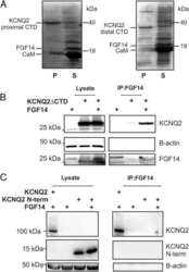
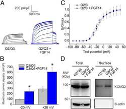
FGF14 is a regulator of KCNQ2/3 channels.
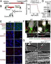
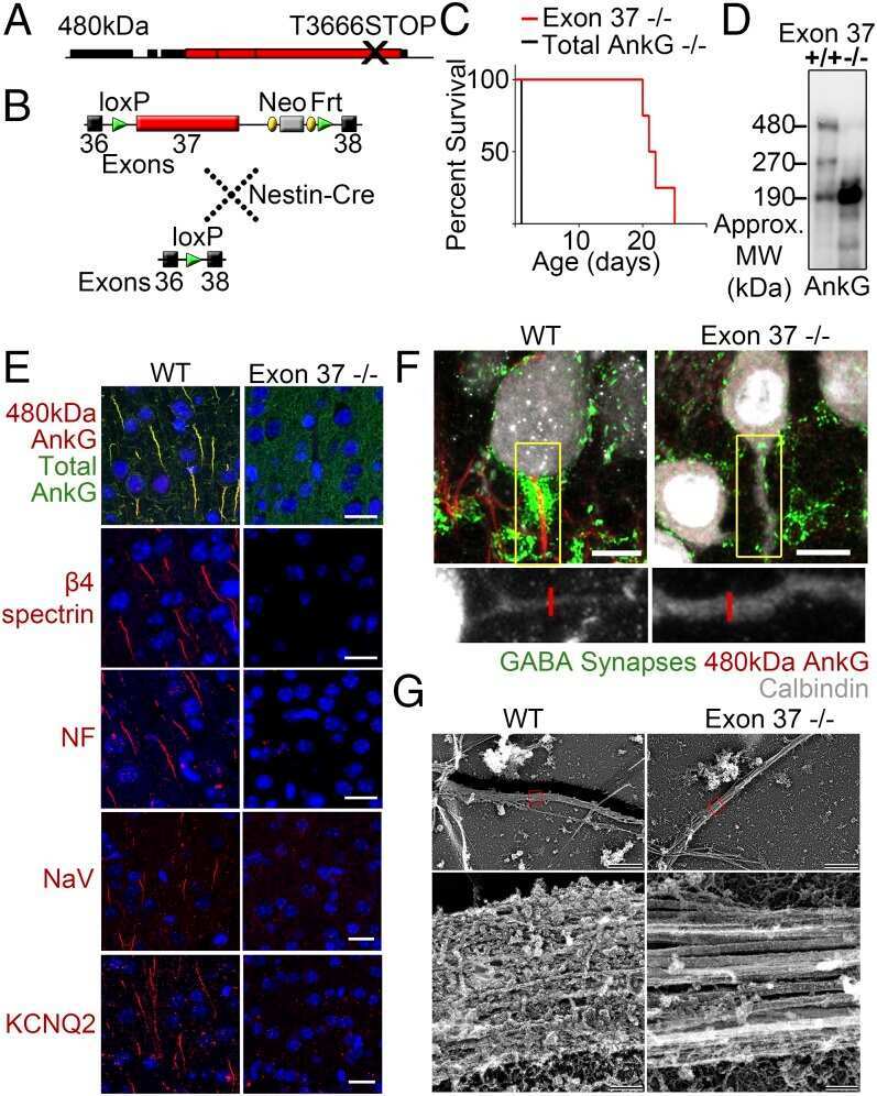
Giant ankyrin-G: a critical innovation in vertebrate evolution of fast and integrated neuronal signaling.
Requirement of subunit co-assembly and ankyrin-G for M-channel localization at the axon initial segment.
Antibodies and a cysteine-modifying reagent show correspondence of M current in neurons to KCNQ2 and KCNQ3 K+ channels.
Nickerson AJ, Rajendran VM
American journal of physiology. Gastrointestinal and liver physiology 2021 Aug 1;321(2):G185-G199
American journal of physiology. Gastrointestinal and liver physiology 2021 Aug 1;321(2):G185-G199
The M-current works in tandem with the persistent sodium current to set the speed of locomotion.
Verneuil J, Brocard C, Trouplin V, Villard L, Peyronnet-Roux J, Brocard F
PLoS biology 2020 Nov;18(11):e3000738
PLoS biology 2020 Nov;18(11):e3000738
FGF14 is a regulator of KCNQ2/3 channels.
Pablo JL, Pitt GS
Proceedings of the National Academy of Sciences of the United States of America 2017 Jan 3;114(1):154-159
Proceedings of the National Academy of Sciences of the United States of America 2017 Jan 3;114(1):154-159
Giant ankyrin-G: a critical innovation in vertebrate evolution of fast and integrated neuronal signaling.
Jenkins PM, Kim N, Jones SL, Tseng WC, Svitkina TM, Yin HH, Bennett V
Proceedings of the National Academy of Sciences of the United States of America 2015 Jan 27;112(4):957-64
Proceedings of the National Academy of Sciences of the United States of America 2015 Jan 27;112(4):957-64
Requirement of subunit co-assembly and ankyrin-G for M-channel localization at the axon initial segment.
Rasmussen HB, Frøkjaer-Jensen C, Jensen CS, Jensen HS, Jørgensen NK, Misonou H, Trimmer JS, Olesen SP, Schmitt N
Journal of cell science 2007 Mar 15;120(Pt 6):953-63
Journal of cell science 2007 Mar 15;120(Pt 6):953-63
Antibodies and a cysteine-modifying reagent show correspondence of M current in neurons to KCNQ2 and KCNQ3 K+ channels.
Roche JP, Westenbroek R, Sorom AJ, Hille B, Mackie K, Shapiro MS
British journal of pharmacology 2002 Dec;137(8):1173-86
British journal of pharmacology 2002 Dec;137(8):1173-86
No comments: Submit comment
Supportive validation
- Submitted by
- Invitrogen Antibodies (provider)
- Main image

- Experimental details
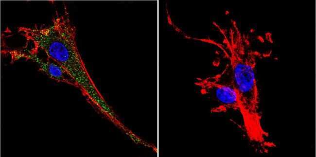
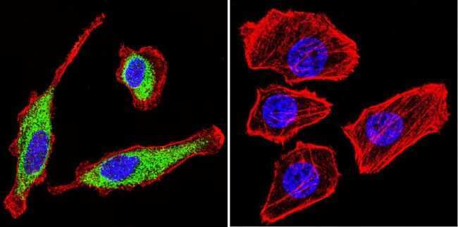
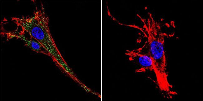
- Immunofluorescent analysis of KCNQ2 using Anti-KCNQ2 Polyclonal Antibody (Product # PA1-929) shows staining in C6 Cells. KCNQ2 staining (green), F-Actin staining with Phalloidin (red) and nuclei with DAPI (blue) is shown. Cells were grown on chamber slides and fixed with formaldehyde prior to staining. Cells were probed without (control) or with or an antibody recognizing KCNQ2 (Product # PA1-929) at a dilution of 1:100 over night at 4°C, washed with PBS and incubated with a DyLight-488 conjugated secondary antibody (Product # 35552, Goat Anti-Rabbit). Images were taken at 60X magnification.
- Submitted by
- Invitrogen Antibodies (provider)
- Main image

- Experimental details
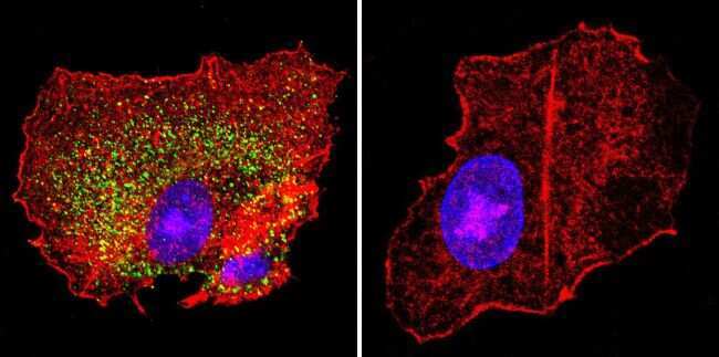
- Immunofluorescent analysis of KCNQ2 using Anti-KCNQ2 Polyclonal Antibody (Product # PA1-929) shows staining in HepG2 Cells. KCNQ2 staining (green), F-Actin staining with Phalloidin (red) and nuclei with DAPI (blue) is shown. Cells were grown on chamber slides and fixed with formaldehyde prior to staining. Cells were probed without (control) or with or an antibody recognizing KCNQ2 (Product # PA1-929) at a dilution of 1:100 over night at 4°C, washed with PBS and incubated with a DyLight-488 conjugated secondary antibody (Product # 35552, Goat Anti-Rabbit). Images were taken at 60X magnification.
- Submitted by
- Invitrogen Antibodies (provider)
- Main image
- Experimental details
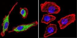
- Immunofluorescent analysis of KCNQ2 using Anti-KCNQ2 Polyclonal Antibody (Product # PA1-929) shows staining in U251 Cells. KCNQ2 staining (green), F-Actin staining with Phalloidin (red) and nuclei with DAPI (blue) is shown. Cells were grown on chamber slides and fixed with formaldehyde prior to staining. Cells were probed without (control) or with or an antibody recognizing KCNQ2 (Product # PA1-929) at a dilution of 1:20 over night at 4°C, washed with PBS and incubated with a DyLight-488 conjugated secondary antibody (Product # 35552, Goat Anti-Rabbit). Images were taken at 60X magnification.
- Submitted by
- Invitrogen Antibodies (provider)
- Main image

- Experimental details
- Immunofluorescent analysis of KCNQ2 using Anti-KCNQ2 Polyclonal Antibody (Product # PA1-929) shows staining in C6 Cells. KCNQ2 staining (green), F-Actin staining with Phalloidin (red) and nuclei with DAPI (blue) is shown. Cells were grown on chamber slides and fixed with formaldehyde prior to staining. Cells were probed without (control) or with or an antibody recognizing KCNQ2 (Product # PA1-929) at a dilution of 1:100 over night at 4°C, washed with PBS and incubated with a DyLight-488 conjugated secondary antibody (Product # 35552, Goat Anti-Rabbit). Images were taken at 60X magnification.
- Submitted by
- Invitrogen Antibodies (provider)
- Main image

- Experimental details
- Immunofluorescent analysis of KCNQ2 using Anti-KCNQ2 Polyclonal Antibody (Product # PA1-929) shows staining in HepG2 Cells. KCNQ2 staining (green), F-Actin staining with Phalloidin (red) and nuclei with DAPI (blue) is shown. Cells were grown on chamber slides and fixed with formaldehyde prior to staining. Cells were probed without (control) or with or an antibody recognizing KCNQ2 (Product # PA1-929) at a dilution of 1:100 over night at 4°C, washed with PBS and incubated with a DyLight-488 conjugated secondary antibody (Product # 35552, Goat Anti-Rabbit). Images were taken at 60X magnification.

- Submitted by
- Invitrogen Antibodies (provider)
- Main image

- Experimental details
- Immunofluorescent analysis of KCNQ2 using Anti-KCNQ2 Polyclonal Antibody (Product # PA1-929) shows staining in U251 Cells. KCNQ2 staining (green), F-Actin staining with Phalloidin (red) and nuclei with DAPI (blue) is shown. Cells were grown on chamber slides and fixed with formaldehyde prior to staining. Cells were probed without (control) or with or an antibody recognizing KCNQ2 (Product # PA1-929) at a dilution of 1:20 over night at 4°C, washed with PBS and incubated with a DyLight-488 conjugated secondary antibody (Product # 35552, Goat Anti-Rabbit). Images were taken at 60X magnification.

Supportive validation
- Submitted by
- Invitrogen Antibodies (provider)
- Main image
- Experimental details
- NULL
- Submitted by
- Invitrogen Antibodies (provider)
- Main image
- Experimental details
- NULL

- Submitted by
- Invitrogen Antibodies (provider)
- Main image
- Experimental details
- NULL

- Submitted by
- Invitrogen Antibodies (provider)
- Main image
- Experimental details
- NULL
- Submitted by
- Invitrogen Antibodies (provider)
- Main image
- Experimental details
- NULL
- Submitted by
- Invitrogen Antibodies (provider)
- Main image
- Experimental details
- NULL
- Submitted by
- Invitrogen Antibodies (provider)
- Main image
- Experimental details
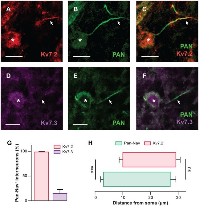
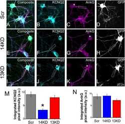
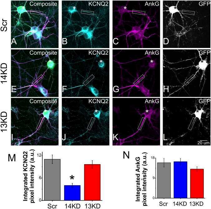
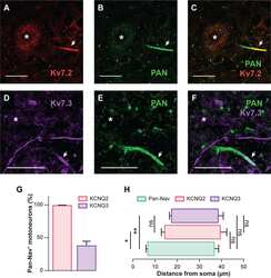
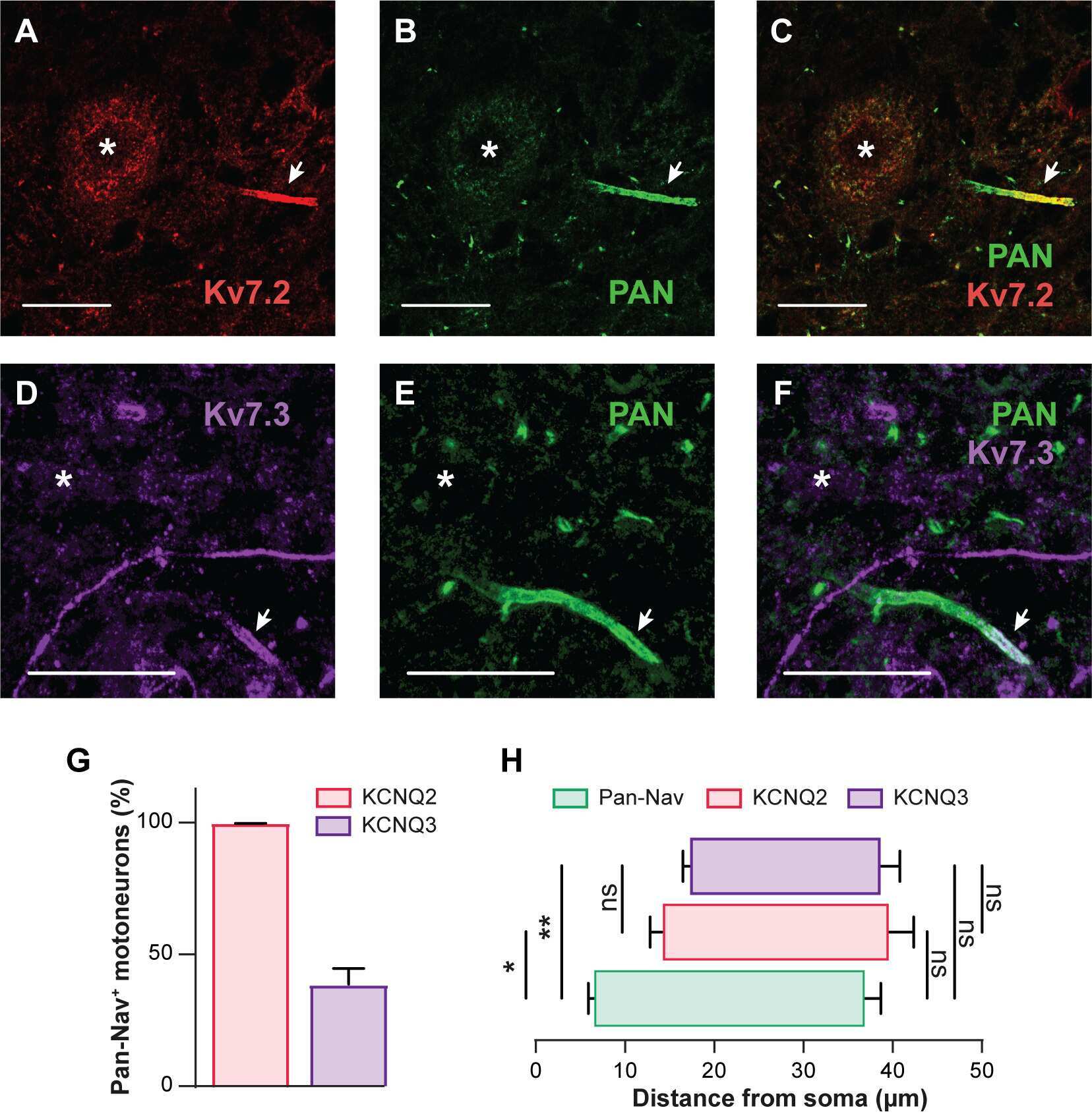
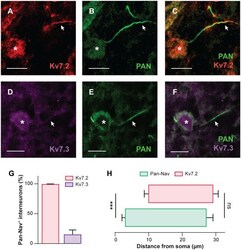
- Fig 2 Lumbar interneurons within the locomotor CPG region ubiquitously express Kv7.2-containing channels. (A-F) Immunostaining of lumbar (L1-L2) ventromedial interneurons from juvenile rats ( n = 3 rats) against Kv7.2 (A, n = 216 cells) or Kv7.3 (D, n = 170 cells) along the AIS labeled by the pan-Na v antibody (B,E). Kv7.2 and pan-Na v are merged in (C), and Kv7.3 and pan-Na v are merged in (F). Asterisks indicate the nucleus position and arrowheads the AIS. Scale bars = 10 mum. (G) Group means quantification of the proportion of pan-Na v positive interneurons expressing Kv7.2 or Kv7.3 channels. (H) Group means quantification of the start and end positions of pan-Na v and Kv7.2 immunolabeling along the axonal process from the soma ( n = 10 cells). *** P < 0.001, comparing start or end positions between groups; Mann-Whitney test. Data are mean +- SEM. Underlying numerical values can be found in the S1 Data . AIS, axonal initial segment; CPG, central pattern generator; Na v , voltage-gated sodium channel.