 Explore
 Validate
 Learn
LearnNBP1-03363
antibody from Novus Biologicals
Targeting: LPAR1
edg-2, EDG2, Gpcr26, GPR26, LPA1, Mrec1.3, rec.1.3, vzg-1
 Western blot
 Immunocytochemistry
Antibody data
- Antibody Data
- Antigen structure
- References [3]
- Comments [0]
- Validations
- Western blot [2]
- Immunohistochemistry [1]
Submit
Validation data
Reference
Comment
Report error
- Product number
- NBP1-03363 - Provider product page

- Provider
- Novus Biologicals
- Proper citation
- Novus Cat#NBP1-03363, RRID:AB_1520803
- Product name
- Rabbit Polyclonal LPAR1/LPA1/EDG-2 Antibody
- Antibody type
- Polyclonal
- Description
- Immunogen affinity purified.
- Reactivity
- Human, Mouse, Rat, Chicken/Avian
- Host
- Rabbit
- Isotype
- IgG
- Vial size
- 0.1 ml
- Concentration
- 1 mg/ml
- Storage
- Store at 4C short term. Aliquot and store at -20C long term. Avoid freeze-thaw cycles.
Submitted references Rho/ROCK acts downstream of lysophosphatidic acid receptor 1 in modulating P2X3 receptor-mediated bone cancer pain in rats.
Massively parallel sequencing reveals an accumulation of de novo mutations and an activating mutation of LPAR1 in a patient with metastatic neuroblastoma.
Primary human endothelial cells secrete agents that reduce responsiveness to lysophosphatidic acid (LPA).
Wu JX, Yuan XM, Wang Q, Wei W, Xu MY
Molecular pain 2016;12
Molecular pain 2016;12
Massively parallel sequencing reveals an accumulation of de novo mutations and an activating mutation of LPAR1 in a patient with metastatic neuroblastoma.
Wei JS, Johansson P, Chen L, Song YK, Tolman C, Li S, Hurd L, Patidar R, Wen X, Badgett TC, Cheuk AT, Marshall JC, Steeg PS, Vaqué Díez JP, Yu Y, Gutkind JS, Khan J
PloS one 2013;8(10):e77731
PloS one 2013;8(10):e77731
Primary human endothelial cells secrete agents that reduce responsiveness to lysophosphatidic acid (LPA).
Park EY, Kazlauskas A
Bioscience reports 2012 Aug;32(4):393-400
Bioscience reports 2012 Aug;32(4):393-400
No comments: Submit comment
Supportive validation
- Submitted by
- Novus Biologicals (provider)
- Main image

- Experimental details
- Simple Western: LPAR1/LPA1/EDG-2 Antibody [NBP1-03363] - Simple Western lane view shows a specific band for EDG2 in 0.5 mg/ml of A431 lysate. This experiment was performed under reducing conditions using the 12-230 kDa separation system.

- Submitted by
- Novus Biologicals (provider)
- Main image
- Experimental details
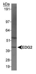
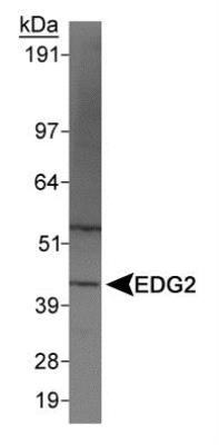
- Western Blot: LPAR1/LPA1/EDG-2 Antibody [NBP1-03363] - Detection of EDG2 in A549 whole cell extracts using NBP1-03363. The band at ~41 kDa position represents the target protein EDG2, whereas, the band at ~55kDa may potentially be the post-translationally modified (glycosylated, palmitoylated or lipidated) form of this protein.
Supportive validation
- Submitted by
- Novus Biologicals (provider)
- Main image
- Experimental details
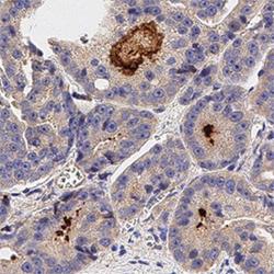
- Immunohistochemistry-Paraffin: LPAR1/LPA1/EDG-2 Antibody [NBP1-03363] - IHC analysis of formalin fixed paraffin-embedded (FFPE) human prostate cancer using LPAR1 antibody at 1:100 on a Bond Rx autostainer (Leica Biosystems). The assay involved 20 minutes of heat induced antigen retrieval (HIER) using 10mM sodium citrate buffer (pH 6.0) and endogenous peroxidase quenching with peroxide block. The sections were incubated with primary antibody for 30 minutes and Bond Polymer Refine Detection (Leica Biosystems) with DAB was used for signal development followed by counterstaining with hematoxylin. Whole slide scanning and capturing of representative images was performed using Aperio AT2 (Leica Biosystems). Staining was obseved in the scattered ducts. Staining was performed by Histowiz.
