Explore
 Validate
 Learn
Learn Western blot
Antibody data
- Antibody Data
- Antigen structure
- References [0]
- Comments [0]
- Validations
- Western blot [2]
- Immunohistochemistry [1]
Submit
Validation data
Reference
Comment
Report error
- Product number
- PA5-52323 - Provider product page

- Provider
- Invitrogen Antibodies
- Product name
- Neuroligin 1 Polyclonal Antibody
- Antibody type
- Polyclonal
- Antigen
- Recombinant full-length protein
- Description
- Immunogen sequence: DQLYLHIGLK PRVKEHYRAN KVNLWLELVP HLHNLNDISQ YTSTTTKVPS TDITFRPTRK NSVPVTSAFP TAKQDDPKQQ PSPFSVDQRD YS Highest antigen sequence identity to the following orthologs: Mouse - 98%, Rat - 98%.
- Reactivity
- Human
- Host
- Rabbit
- Isotype
- IgG
- Vial size
- 100 µL
- Concentration
- 0.1 mg/mL
- Storage
- Store at 4°C short term. For long term storage, store at -20°C, avoiding freeze/thaw cycles.
No comments: Submit comment
Supportive validation
- Submitted by
- Invitrogen Antibodies (provider)
- Main image

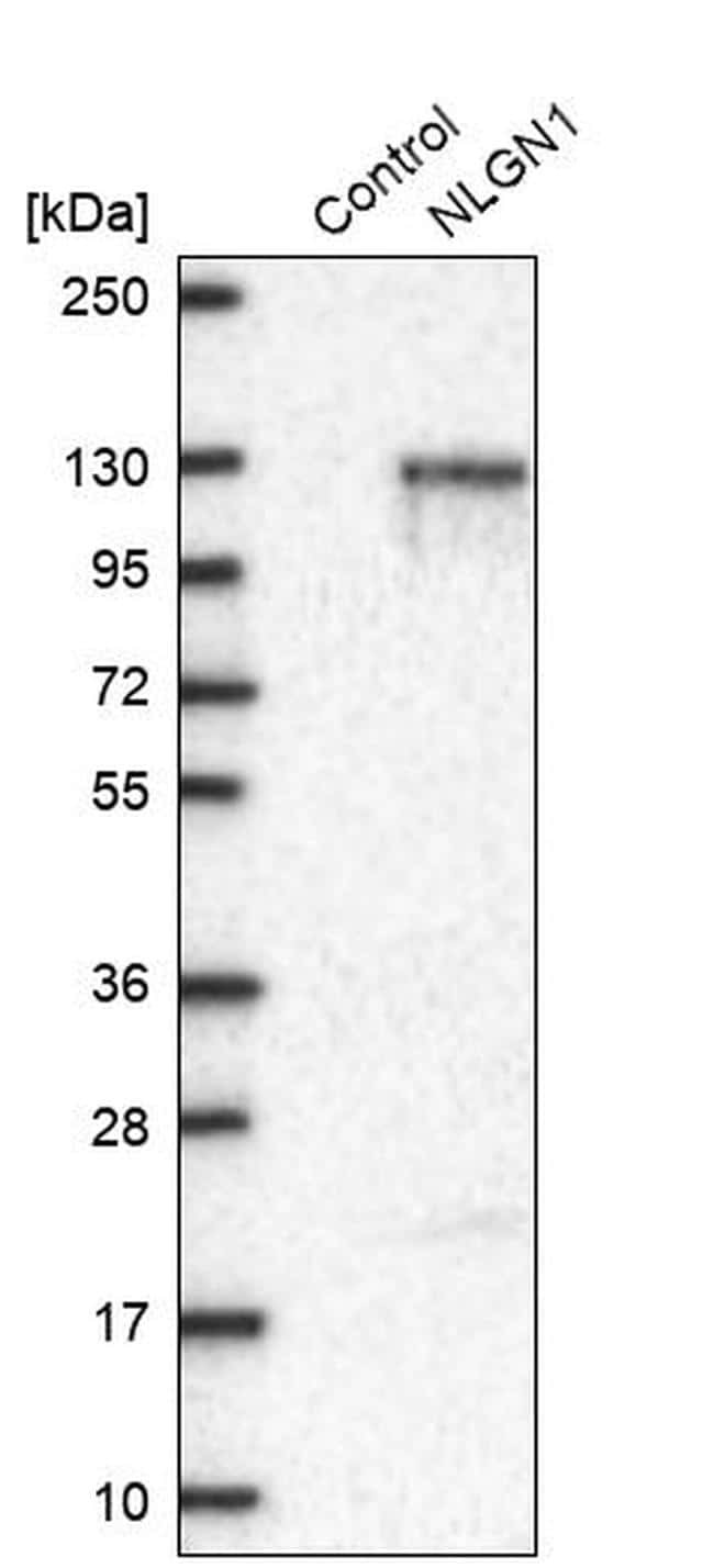
- Experimental details
- Western blot analysis of Neuroligin 1 in control (vector only transfected HEK293T lysate) and NLGN1 over-expression lysate (Co-expressed with a C-terminal myc-DDK tag (~3.1 kDa) in mammalian HEK293T cells). Samples were probed using a Neuroligin 1 Polyclonal Antibody (Product # PA5-52323).
- Submitted by
- Invitrogen Antibodies (provider)
- Main image
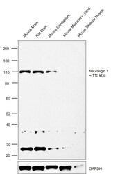
- Experimental details
- Western Blot was performed using Anti-Neuroligin 1 Polyclonal Antibody (Product # PA5-52323) and a 110 kDa band corresponding to Neuroligin-1 was observed only in Mouse Brain, Rat Brain and Mouse Cerebellum which are reported to be positive and not in any other tissues like Mouse Mammary gland and Skeletal muscle. Tissue extracts (30 µg lysate) of Mouse Brain (Lane 1), Rat Brain (Lane 2), Mouse Cerebellum (Lane 3), Mouse Mammary gland (Lane 4) and Mouse Skeletal Muscle (Lane 5) were electrophoresed using NuPAGE™ 4-12% Bis-Tris Protein Gel (Product # NP0322BOX). Resolved proteins were then transferred onto a Nitrocellulose membrane (Product # IB23001) by iBlot® 2 Dry Blotting System (Product # IB21001). The Blot was probed with the primary antibody (0.2 µg/mL) and detected by chemiluminescence with Goat anti-Rabbit IgG (H+L) Superclonal™ Recombinant Secondary Antibody, HRP (Product # A27036, 1:4000 dilution) using the iBright FL 1000 (Product # A32752). Chemiluminescent detection was performed using SuperSignal™ West Dura Extended Duration Substrate (Product # 34076).An uncharacterized band of ~25 kDa and 35 kDa were also observed across the tissues.

Supportive validation
- Submitted by
- Invitrogen Antibodies (provider)
- Main image
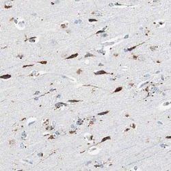
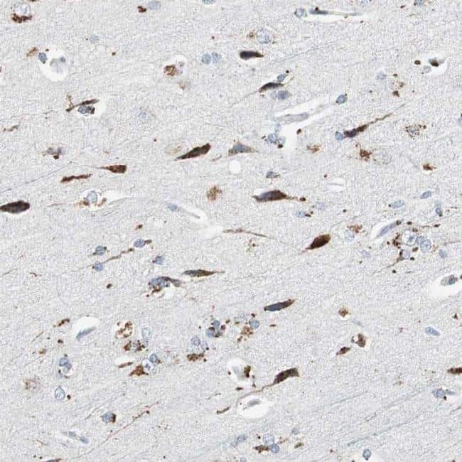
- Experimental details
- Immunohistochemical staining of NLGN1 in human cerebral cortex tissue shows strong cytoplasmic positivity in neuronal cells. Samples were probed using a NLGN1 Polyclonal Antibody (Product # PA5-52323).