Explore
 Validate
 Learn
Learn Western blot
 Immunocytochemistry
Antibody data
- Antibody Data
- Antigen structure
- References [2]
- Comments [0]
- Validations
- Immunocytochemistry [2]
- Immunoprecipitation [1]
- Immunohistochemistry [7]
- Other assay [3]
Submit
Validation data
Reference
Comment
Report error
- Product number
- PA5-31101 - Provider product page

- Provider
- Invitrogen Antibodies
- Product name
- eIF4G3 Polyclonal Antibody
- Antibody type
- Polyclonal
- Antigen
- Recombinant full-length protein
- Description
- Recommended positive controls: 293T, A431, HeLa, HepG2, mouse testis. Predicted reactivity: Human (99%), Mouse (96%), Rat (97%). Store product as a concentrated solution. Centrifuge briefly prior to opening the vial.
- Reactivity
- Human, Mouse, Rat
- Host
- Rabbit
- Isotype
- IgG
- Vial size
- 100 μL
- Concentration
- 0.26 mg/mL
- Storage
- Store at 4°C short term. For long term storage, store at -20°C, avoiding freeze/thaw cycles.
Submitted references eIF4E3 forms an active eIF4F complex during stresses (eIF4FS) targeting mTOR and re-programs the translatome.
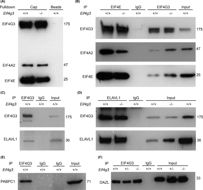
Nuclear localization of EIF4G3 suggests a role for the XY body in translational regulation during spermatogenesis in mice.
Weiss B, Allen GE, Kloehn J, Abid K, Jaquier-Gubler P, Curran JA
Nucleic acids research 2021 May 21;49(9):5159-5176
Nucleic acids research 2021 May 21;49(9):5159-5176
Nuclear localization of EIF4G3 suggests a role for the XY body in translational regulation during spermatogenesis in mice.
Hu J, Sun F, Handel MA
Biology of reproduction 2018 Jan 1;98(1):102-114
Biology of reproduction 2018 Jan 1;98(1):102-114
No comments: Submit comment
Supportive validation
- Submitted by
- Invitrogen Antibodies (provider)
- Main image
- Experimental details
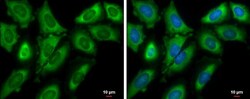
- Immunocytochemistry-Immunofluorescence analysis of eIF4G3 was performed in HeLa cells fixed in 4% paraformaldehyde at RT for 15 min. Green: eIF4G3 Polyclonal Antibody (Product # PA5-31101) diluted at 1:500. Blue: Hoechst 33342 staining. Scale bar = 10 µm.

- Submitted by
- Invitrogen Antibodies (provider)
- Main image

- Experimental details
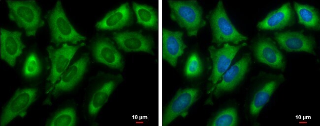
- Immunocytochemistry-Immunofluorescence analysis of eIF4G3 was performed in HeLa cells fixed in 4% paraformaldehyde at RT for 15 min. Green: eIF4G3 Polyclonal Antibody (Product # PA5-31101) diluted at 1:500. Blue: Hoechst 33342 staining. Scale bar = 10 µm.
Supportive validation
- Submitted by
- Invitrogen Antibodies (provider)
- Main image
- Experimental details
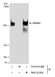
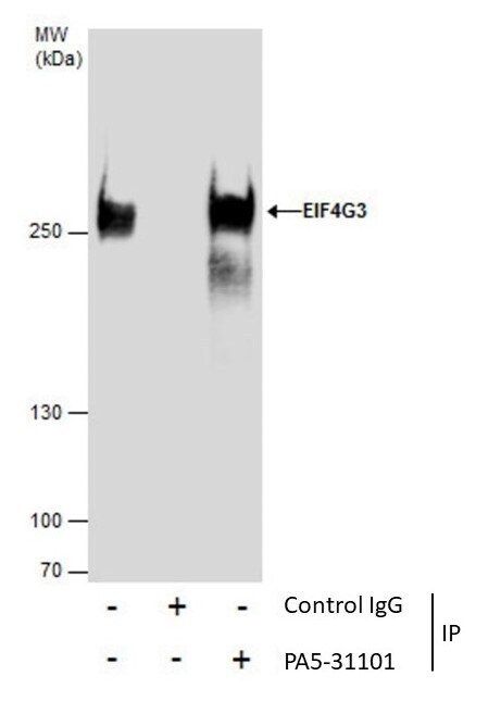
- Immunoprecipitation of EIF4G3 was performed in 293T whole cell extracts using 5 µg of eIF4G3 Polyclonal Antibody (Product # PA5-31101). Samples were transferred to a membrane and probed with eIF4G3 Polyclonal Antibody as a primary antibody and an HRP-conjugated anti-Rabbit IgG was used as a secondary antibody.

Supportive validation
- Submitted by
- Invitrogen Antibodies (provider)
- Main image
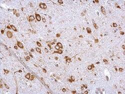
- Experimental details
- eIF4G3 Polyclonal Antibody detects EIF4G3 protein at cytosol on mouse brain stem by immunohistochemical analysis. Sample: Paraffin-embedded mouse brain stem. EIF4G3 Polyclonal Antibody (Product # PA5-31101) dilution: 1:500. Antigen Retrieval: EDTA based buffer, pH 8.0, 15 min.
- Submitted by
- Invitrogen Antibodies (provider)
- Main image
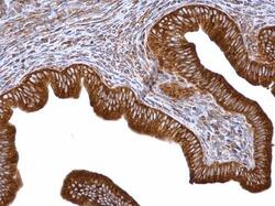
- Experimental details
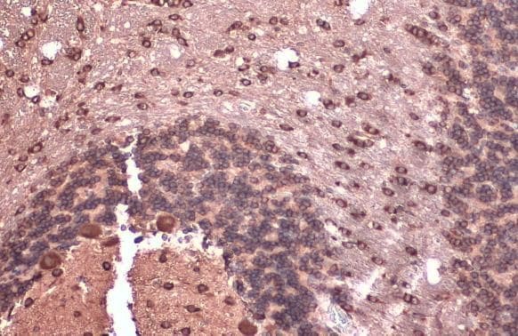
- eIF4G3 Polyclonal Antibody detects EIF4G3 protein at cytosol on mouse cervix by immunohistochemical analysis. Sample: Paraffin-embedded mouse cervix. EIF4G3 Polyclonal Antibody (Product # PA5-31101) dilution: 1:500. Antigen Retrieval: EDTA based buffer, pH 8.0, 15 min.
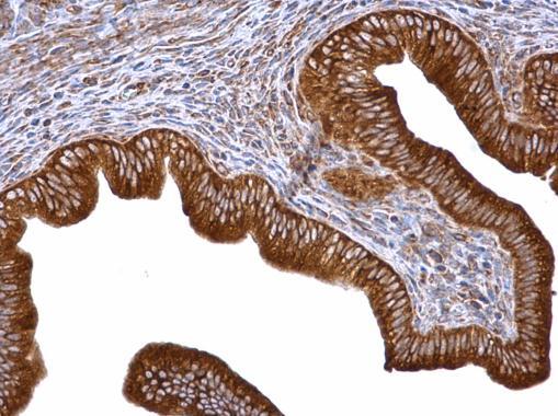
- Submitted by
- Invitrogen Antibodies (provider)
- Main image
- Experimental details
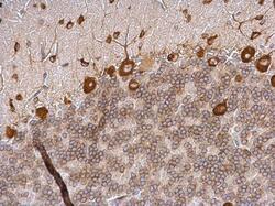
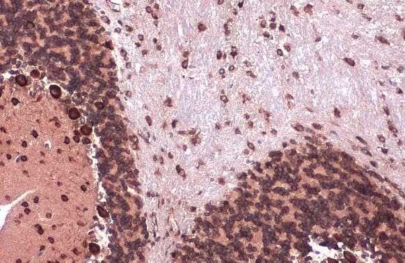
- eIF4G3 Polyclonal Antibody detects EIF4G3 protein at cytosol on rat hind brain by immunohistochemical analysis. Sample: Paraffin-embedded rat hind brain. EIF4G3 Polyclonal Antibody (Product # PA5-31101) dilution: 1:500. Antigen Retrieval: EDTA based buffer, pH 8.0, 15 min.

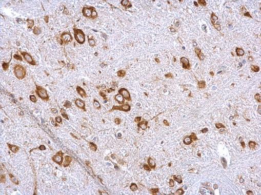
- Submitted by
- Invitrogen Antibodies (provider)
- Main image
- Experimental details
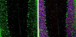
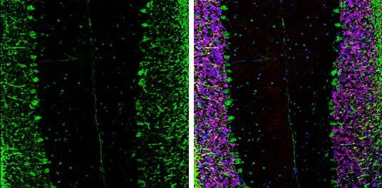
- Immunohistochemistry (Frozen) analysis of eIF4G3 was performed in frozen-sectioned adult mouse cerebellum tissue using eIF4G3 Polyclonal Antibody (Product # PA5-31101) at a dilution of 1:250 (Green). Red: NeuN, stained by NeuN antibody diluted at 1:500. Blue: Fluoroshield with DAPI.
- Submitted by
- Invitrogen Antibodies (provider)
- Main image
- Experimental details
- Immunohistochemistry (Paraffin) analysis of eIF4G3 was performed in paraffin-embedded mouse brain tissue using eIF4G3 Polyclonal Antibody (Product # PA5-31101) at a dilution of 1:500. Antigen Retrieval: Citrate buffer, pH 6.0, 15 min.
- Submitted by
- Invitrogen Antibodies (provider)
- Main image
- Experimental details
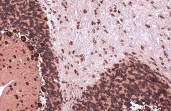
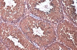
- Immunohistochemistry (Paraffin) analysis of eIF4G3 was performed in paraffin-embedded mouse testis tissue using eIF4G3 Polyclonal Antibody (Product # PA5-31101) at a dilution of 1:500. Antigen Retrieval: Citrate buffer, pH 6.0, 15 min.

- Submitted by
- Invitrogen Antibodies (provider)
- Main image
- Experimental details
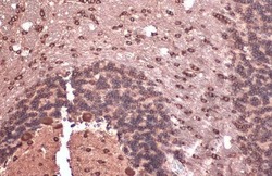
- Immunohistochemistry (Paraffin) analysis of eIF4G3 was performed in paraffin-embedded rat brain tissue using eIF4G3 Polyclonal Antibody (Product # PA5-31101) at a dilution of 1:500. Antigen Retrieval: Citrate buffer, pH 6.0, 15 min.
Supportive validation
- Submitted by
- Invitrogen Antibodies (provider)
- Main image

- Experimental details
- NULL
- Submitted by
- Invitrogen Antibodies (provider)
- Main image

- Experimental details
- Immunoprecipitation of EIF4G3 was performed in 293T whole cell extracts using 5 µg of eIF4G3 Polyclonal Antibody (Product # PA5-31101). Samples were transferred to a membrane and probed with eIF4G3 Polyclonal Antibody as a primary antibody and an HRP-conjugated anti-Rabbit IgG was used as a secondary antibody.
- Submitted by
- Invitrogen Antibodies (provider)
- Main image
- Experimental details
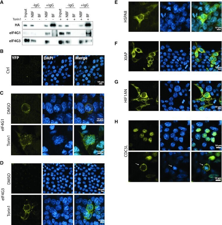
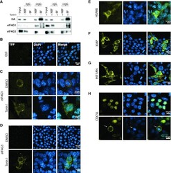
- Figure 3. The eIF4E3 partners discovered by Y2H. ( A ) Western blot analysis of the co-IP assay using eIF4E3 HA and endogenous eIF4G1 or eIF4G3 starting from a lysate of HEK293T cells transduced with eIF4E3 HA treated with DMSO or with 250 nM Torin1 for 2 h. Beads carrying covalently cross-linked Anti-HA Ab (+IgG) were used to immunoprecipitate eIF4E3 HA . Beads without Ab served as a control (-IgG). (B-H) Bimolecular complementation assay using Venus YFP in transfected HEK293T cells. Images were generated by confocal microscopy. Venus fragment 1 and Venus fragment 2 were fused to eIF4E3 and to one of the tested partners, respectively: empty vector ( B ), eIF4G1 ( C ), eIF4G3 ( D ), HSPA8 ( E ), XIAP ( F ), HIF1AN ( G ) and CDC5L ( H ).