Explore
 Validate
 Learn
Learn Western blot
Antibody data
- Antibody Data
- Antigen structure
- References [1]
- Comments [0]
- Validations
- Western blot [4]
- Immunocytochemistry [3]
- Immunoprecipitation [1]
- Immunohistochemistry [4]
Submit
Validation data
Reference
Comment
Report error
- Product number
- GTX118109 - Provider product page

- Provider
- GeneTex
- Proper citation
- GeneTex Cat#GTX118109, RRID:AB_11167995
- Product name
- EIF4G3 antibody [N1], N-term
- Antibody type
- Polyclonal
- Reactivity
- Human, Mouse, Rat
- Host
- Rabbit
Submitted references Resources for the Comprehensive Discovery of Functional RNA Elements.
Sundararaman B, Zhan L, Blue SM, Stanton R, Elkins K, Olson S, Wei X, Van Nostrand EL, Pratt GA, Huelga SC, Smalec BM, Wang X, Hong EL, Davidson JM, Lécuyer E, Graveley BR, Yeo GW
Molecular cell 2016 Mar 17;61(6):903-13
Molecular cell 2016 Mar 17;61(6):903-13
No comments: Submit comment
Supportive validation
- Submitted by
- GeneTex (provider)
- Main image

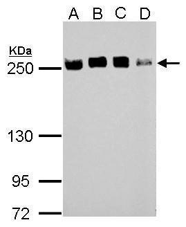
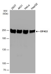
- Experimental details
- Sample (30 ?g of whole cell lysate) A: 293T B: A431 C: HeLa D: HepG2 5% SDS PAGE GTX118109 diluted at 1:10000 The HRP-conjugated anti-rabbit IgG antibody (GTX213110-01) was used to detect the primary antibody.
- Submitted by
- GeneTex (provider)
- Main image

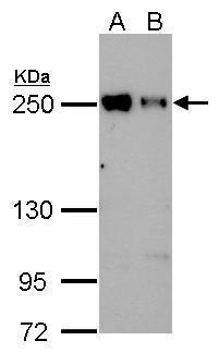
- Experimental details
- Sample (30 ?g of whole cell lysate) A: JC B: BCL-1 5% SDS PAGE GTX118109 diluted at 1:5000 The HRP-conjugated anti-rabbit IgG antibody (GTX213110-01) was used to detect the primary antibody.
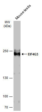
- Submitted by
- GeneTex (provider)
- Main image
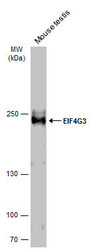
- Experimental details
- Mouse tissue extract (50 ?g) was separated by 5% SDS-PAGE, and the membrane was blotted with EIF4G3 antibody [N1], N-term (GTX118109) diluted at 1:1000. The HRP-conjugated anti-rabbit IgG antibody (GTX213110-01) was used to detect the primary antibody.
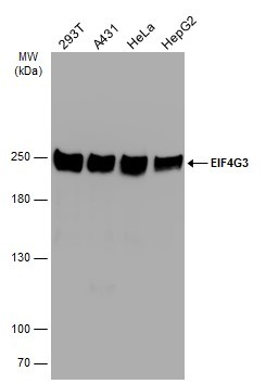
- Submitted by
- GeneTex (provider)
- Main image
- Experimental details
- Various whole cell extracts (30 ?g) were separated by 5% SDS-PAGE, and the membrane was blotted with EIF4G3 antibody [N1], N-term (GTX118109) diluted at 1:10000.
Supportive validation
- Submitted by
- GeneTex (provider)
- Main image
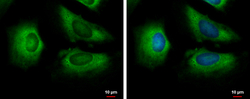
- Experimental details
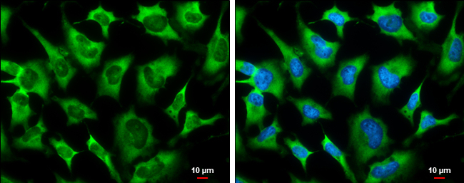
- EIF4G3 antibody [N1], N-term detects EIF4G3 protein at cytoplasm by immunofluorescent analysis.Sample: HeLa cells were fixed in 4% paraformaldehyde at RT for 15 min.Green: EIF4G3 protein stained by EIF4G3 antibody [N1], N-term (GTX118109) diluted at 1:500.Blue: Hoechst 33342 staining.

- Submitted by
- GeneTex (provider)
- Main image

- Experimental details
- EIF4G3 antibody [N1], N-term detects EIF4G3 protein at cytoplasm by immunofluorescent analysis.Sample: HeLa cells were fixed in 4% paraformaldehyde at RT for 15 min.Green: EIF4G3 protein stained by EIF4G3 antibody [N1], N-term (GTX118109) diluted at 1:500.Blue: Hoechst 33342 staining.Scale bar = 10 £gm.
- Submitted by
- GeneTex (provider)
- Main image

- Experimental details
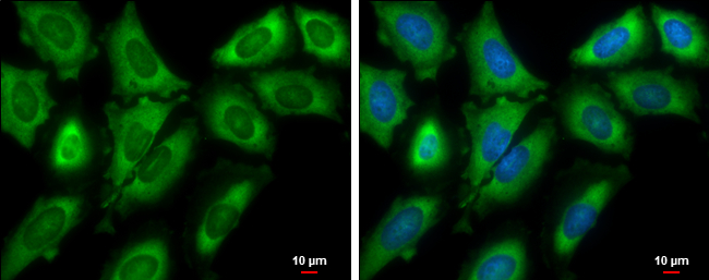
- EIF4G3 antibody [N1], N-term detects EIF4G3 protein at cytoplasm by immunofluorescent analysis.Sample: HeLa cells were fixed in 4% paraformaldehyde at RT for 15 min.Green: EIF4G3 protein stained by EIF4G3 antibody [N1], N-term (GTX118109) diluted at 1:500.Blue: Hoechst 33342 staining.Scale bar = 10 £gm.
Supportive validation
- Submitted by
- GeneTex (provider)
- Main image

- Experimental details
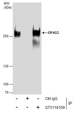
- Immunoprecipitation of EIF4G3 protein from 293T whole cell extracts using 5 £gg of EIF4G3 antibody [N1], N-term (GTX118109).Western blot analysis was performed using EIF4G3 antibody [N1], N-term (GTX118109).EasyBlot anti-Rabbit IgG (GTX221666-01) was used as a secondary reagent.
Supportive validation
- Submitted by
- GeneTex (provider)
- Main image
- Experimental details
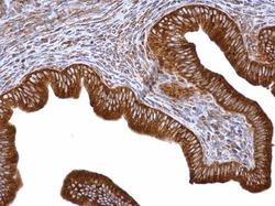
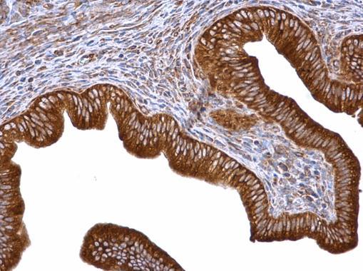
- EIF4G3 antibody [N1], N-term detects EIF4G3 protein at cytosol on mouse cervix by immunohistochemical analysis. Sample: Paraffin-embedded mouse cervix. EIF4G3 antibody [N1], N-term (GTX118109) dilution: 1:500.
- Submitted by
- GeneTex (provider)
- Main image

- Experimental details
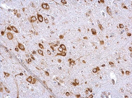
- EIF4G3 antibody [N1], N-term detects EIF4G3 protein at cytosol on rat hind brain by immunohistochemical analysis. Sample: Paraffin-embedded rat hind brain. EIF4G3 antibody [N1], N-term (GTX118109) dilution: 1:500.
- Submitted by
- GeneTex (provider)
- Main image

- Experimental details
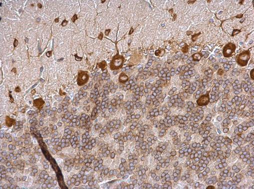
- EIF4G3 antibody [N1], N-term detects EIF4G3 protein at cytosol on mouse brain stem by immunohistochemical analysis. Sample: Paraffin-embedded mouse brain stem. EIF4G3 antibody [N1], N-term (GTX118109) dilution: 1:500.
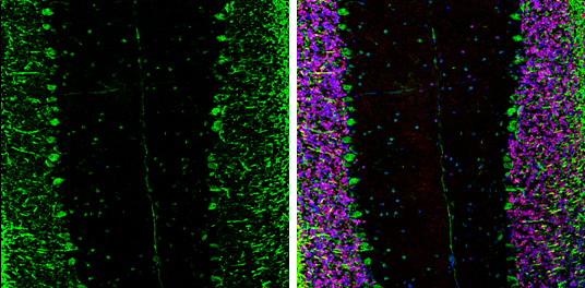
- Submitted by
- GeneTex (provider)
- Main image
- Experimental details
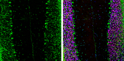
- EIF4G3 antibody [N1], N-term detects EIF4G3 protein expression by immunohistochemical analysis.Sample: Frozen-sectioned adult mouse cerebellum. Green: EIF4G3 protein stained by EIF4G3 antibody [N1], N-term (GTX118109) diluted at 1:250.Red: NeuN, stained by NeuN antibody [2Q158] (GTX30773) diluted at 1:500.Blue: Fluoroshield with DAPI (GTX30920).