 Explore
 Validate
 Learn
Learn Immunocytochemistry
 Immunohistochemistry
Antibody data
- Antibody Data
- Antigen structure
- References [0]
- Comments [0]
- Validations
- Immunohistochemistry [1]
Submit
Validation data
Reference
Comment
Report error
- Product number
- GTX80746 - Provider product page

- Provider
- GeneTex
- Proper citation
- GeneTex Cat#GTX80746, RRID:AB_625998
- Product name
- NTR1 antibody
- Antibody type
- Polyclonal
- Reactivity
- Rat
- Host
- Guinea Pig
No comments: Submit comment
Supportive validation
- Submitted by
- GeneTex (provider)
- Main image
- Experimental details
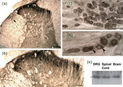
- Expression of NTS1 receptors in sensory neurons and spinal cord. (a) Immunoperoxidase staining reveals the presence of NTS1-like immunoreactivity throughout the lumbar dorsal horn of the spinal cord. Immunolabeling is most prominent in the superficial layers of the dorsal horn. The lateral spinal nucleus (lsn) is also moderately labeled (arrow). (b) High magnification of (a). A dense NTS1 immunostaining is observed over laminae I and II of the dorsal horn. Numerous immunopositive nerve cell bodies are visible in the superficial laminae and in the nucleus proprius of the dorsal horn (arrows). (c) Light microscopic analysis of NTS1 expression in primary afferent neurons. NTS1 is expressed in subpopulations of small- and medium-ganglion cells. No apparent labeling is detected in large DRG neurons (arrows). At higher magnification, NTS1 neurons exhibit a cytoplasmic pattern of immunoreactivity (d, arrowheads). (e) Identification of endogenously expressed NTS1 receptors, by western blotting, in homogenates from DRGs, lumbar spinal cord and brain. The 47 kDa protein band corresponds to the molecular weight deduced from the cDNA sequence of NTS1. Each lane represents the transfer of 25 lg of protein. Scale bars: (a, b, c, and d) 300, 150, 70 and 20 lm, respectively
