Explore
 Validate
 Learn
Learn Western blot
Antibody data
- Antibody Data
- Antigen structure
- References [0]
- Comments [0]
- Validations
- Western blot [1]
- Immunocytochemistry [1]
- Immunohistochemistry [2]
- Flow cytometry [1]
Submit
Validation data
Reference
Comment
Report error
- Product number
- ANT-015-25UL - Provider product page

- Provider
- Invitrogen Antibodies
- Product name
- Neurotensin Receptor 1 (extracellular) Polyclonal Antibody
- Antibody type
- Polyclonal
- Antigen
- Other
- Reactivity
- Human, Rat
- Host
- Rabbit
- Isotype
- IgG
- Vial size
- 25 µL
- Concentration
- 0.8 mg/mL
- Storage
- -20° C, Avoid Freeze/Thaw Cycles
No comments: Submit comment
Supportive validation
- Submitted by
- Invitrogen Antibodies (provider)
- Main image
- Experimental details
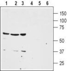
- Western blot analysis of human colon cancer HT-29 (lanes 1 and 4), human lung small cell carcinoma NCI-H526 (lanes 2 and 5) and human breast adenocarcinoma MDA-MB-468 (lanes 3 and 6) cell lysates: - 1-3. Anti-Neurotensin Receptor 1 (extracellular) Antibody (#ANT-015), (1:200).4-6. Anti-Neurotensin Receptor 1 (extracellular) Antibody , preincubated with Neurotensin Receptor 1 (extracellular) Blocking Peptide (#BLP-NT015).
Supportive validation
- Submitted by
- Invitrogen Antibodies (provider)
- Main image
- Experimental details
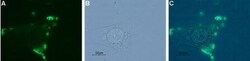
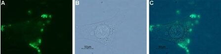
- Expression of NTSR1 in human MCF-7 cells - Cell surface detection of NTSR1 in live intact human MCF-7 breast adenocarcinoma cell line. A. Extracellular staining of cells with Anti-Neurotensin Receptor 1 (extracellular) Antibody (#ANT-015), (1:20), followed by Alexa 488-conjugated goat Anti-rabbit Antibody (green staining). B. Live view of the cells. C. Merge of A and B.
Supportive validation
- Submitted by
- Invitrogen Antibodies (provider)
- Main image
- Experimental details
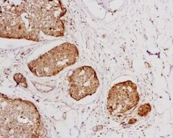
- Expression of NTSR1 in human breast cancer - Immunohistochemical staining ofhuman breast cancer cells using Anti-Neurotensin Receptor 1 (extracellular) Antibody (#ANT-015), (1:100). Staining (brown color) is specific for epithelium-derivate malignant cells. Hematoxilin is used as the counterstain.

- Submitted by
- Invitrogen Antibodies (provider)
- Main image
- Experimental details
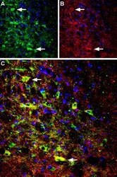
- Expression of NTSR1 in rat brain - Immunohistochemical staining of perfusion-fixed frozen rat brain sections using Anti-Neurotensin Receptor 1 (extracellular) Antibody (#ANT-015), (1:100). A. NTSR1 staining (green) appears in cells of the diagonal band region. B. The same section was stained with Choline acetyltransferase (ChAT) (red), a marker of cholinergic neurons. C. Merge of the two images demonstrates expression of NTSR1 in cholinergic neurons. Arrows point at examples of colocalization. Cell nuclei were stained with DAPI (blue).

Supportive validation
- Submitted by
- Invitrogen Antibodies (provider)
- Main image

- Experimental details
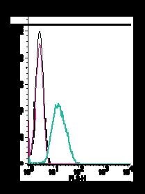
- Cell surface detection of NTSR1 by indirect flow cytometry in live intact human promyelocytic leukemia HL-60 cells: - (black line) cells. (red) Cells + goat- Anti-rabbit-PE. (green) Cells + Anti-Neurotensin Receptor 1 (extracellular) Antibody (#ANT-015), (2.5μg) + goat- Anti-rabbit-PE.