Explore
 Validate
 Learn
Learn Western blot
 Immunocytochemistry
Antibody data
- Antibody Data
- Antigen structure
- References [3]
- Comments [0]
- Validations
- Immunocytochemistry [1]
- Other assay [4]
Submit
Validation data
Reference
Comment
Report error
- Product number
- PA3-214 - Provider product page

- Provider
- Invitrogen Antibodies
- Product name
- NTSR1 Polyclonal Antibody
- Antibody type
- Polyclonal
- Antigen
- Synthetic peptide
- Description
- PA3-214 detects NTSR1 in human samples. PA3-214 has successfully been used in Western blot and immunocytochemical procedures. The PA3-214 immunogen is a synthetic peptide corresponding to the C-terminal amino acids K(398)ADSVSSNHTLSSNATRETLY(409) of NTSR1.
- Reactivity
- Human
- Host
- Rabbit
- Isotype
- IgG
- Vial size
- 100 µL
- Concentration
- Conc. Not Determined
- Storage
- -20° C, Avoid Freeze/Thaw Cycles
Submitted references Neurotensin receptor 1 signaling promotes pancreatic cancer progression.
Neurotensin: A novel mediator of ovulation?
Diverse expression patterns and tumorigenic role of neurotensin signaling components in colorectal cancer cells.
Takahashi K, Ehata S, Miyauchi K, Morishita Y, Miyazawa K, Miyazono K
Molecular oncology 2021 Jan;15(1):151-166
Molecular oncology 2021 Jan;15(1):151-166
Neurotensin: A novel mediator of ovulation?
Campbell GE, Bender HR, Parker GA, Curry TE Jr, Duffy DM
FASEB journal : official publication of the Federation of American Societies for Experimental Biology 2021 Apr;35(4):e21481
FASEB journal : official publication of the Federation of American Societies for Experimental Biology 2021 Apr;35(4):e21481
Diverse expression patterns and tumorigenic role of neurotensin signaling components in colorectal cancer cells.
Kim JT, Weiss HL, Evers BM
International journal of oncology 2017 Jun;50(6):2200-2206
International journal of oncology 2017 Jun;50(6):2200-2206
No comments: Submit comment
Supportive validation
- Submitted by
- Invitrogen Antibodies (provider)
- Main image

- Experimental details
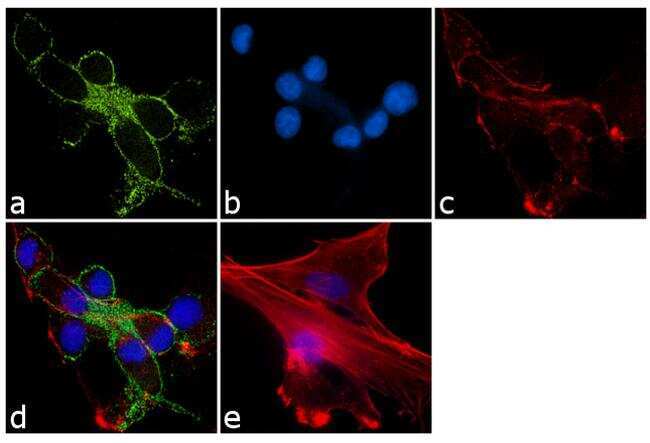
- Immunofluorescence analysis of Neurotensin receptor type 1 (NTSR1) was performed using 90% confluent log phase U-87 MG cells. The cells were fixed with 4% paraformaldehyde for 10 minutes, permeabilized with 0.1% Triton™ X-100 for 10 minutes, and blocked with 1% BSA for 1 hour at room temperature. The cells were labeled with NTSR1 Rabbit Polyclonal Antibody (Product # PA3-214) at 1:250 dilution in 0.1% BSA and incubated for 3 hours at room temperature and then labeled with Goat anti-Rabbit IgG (H+L) Superclonal™ Secondary Antibody, Alexa Fluor® 488 conjugate (Product # A27034) at a dilution of 1:2000 for 45 minutes at room temperature (Panel a: green). Nuclei (Panel b: blue) were stained with SlowFade® Gold Antifade Mountant with DAPI (Product # S36938). F-actin (Panel c: red) was stained with Rhodamine Phalloidin (Product # R415, 1:300). Panel d represents the merged image showing membranous localization. Panel e shows the no primary antibody control. The images were captured at 60X magnification.
Supportive validation
- Submitted by
- Invitrogen Antibodies (provider)
- Main image
- Experimental details
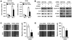
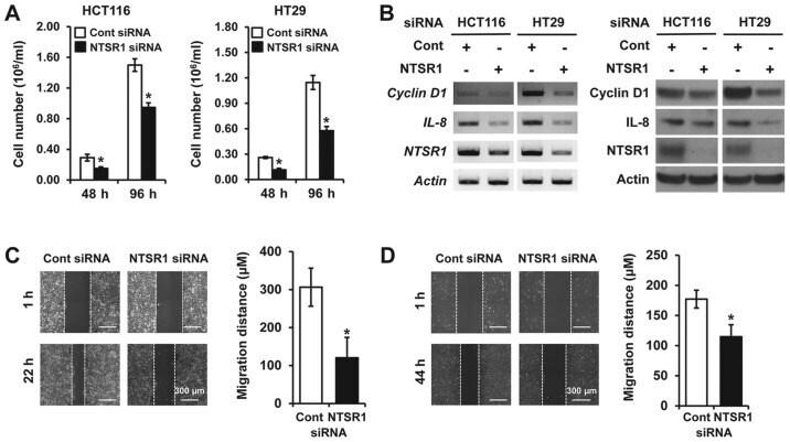
- Figure 3 Silencing of NTSR1 influences cell growth and migration in CRC cells. (A) Equal numbers of HCT116 (left) and HT29 (right) cells transfected with siRNA against non-targeting (Cont) or NTSR1 were seeded in 24-well plates. Cells were counted after 48 and 96 h incubation using a cell counter. * P
- Submitted by
- Invitrogen Antibodies (provider)
- Main image

- Experimental details
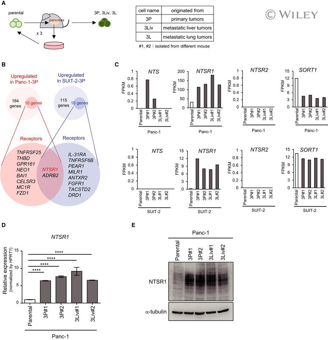
- 1 Fig. Identification of a key molecule for pancreatic cancer, NTSR1, using orthotopic serial transplantation models. (A) Establishment of pancreatic cancer sublines using an orthotopic tumor model [ 14]. Parental Panc-1 cells or parental SUIT-2 cells were inoculated orthotopically into the pancreas in nude mice. Cells were obtained from the primary tumor and then inoculated into another mouse. This process was repeated three times, and highly malignant cancer sublines were established. More than two sublines were established from each tumor model (#1 and #2) and named 3P cells (from the primary tumor), 3Liv cells (from the metastatic liver tumor), and 3L cells (from the metastatic lung tumor). (B) Screening of candidate genes using RNA-seq analysis. The genes were purified as follows: (i) the fragments per kilobase of exon per million mapped sequence reads (FPKM) in 3P cells>3 and (ii) increased more than threefold, compared with the FPKM in parental cells. Among them, genes encoding receptors in both SUIT-2 and Panc-1 cells were purified. The number of purified genes and their receptor gene symbols are shown in the diagram. (C) Expression of NTS and NTSRs in Panc-1 cells (top) and SUIT-2 cells (bottom). Based on RNA-seq analysis, the expression of NTS and NTSRs in Panc-1 cells (parental, 3P, and 3Liv) and SUIT-2 cells (parental, 3P, and 3L) is shown with FPKM. (D) Expression of NTSR1 in Panc-1 cells. Expression of NTSR1 mRNA in pancreatic cancer cells was determined by qRT-
- Submitted by
- Invitrogen Antibodies (provider)
- Main image

- Experimental details
- Fig. 1 Identification of a key molecule for pancreatic cancer, NTSR1, using orthotopic serial transplantation models. (A) Establishment of pancreatic cancer sublines using an orthotopic tumor model [ 14 ]. Parental Panc-1 cells or parental SUIT-2 cells were inoculated orthotopically into the pancreas in nude mice. Cells were obtained from the primary tumor and then inoculated into another mouse. This process was repeated three times, and highly malignant cancer sublines were established. More than two sublines were established from each tumor model (#1 and #2) and named 3P cells (from the primary tumor), 3Liv cells (from the metastatic liver tumor), and 3L cells (from the metastatic lung tumor). (B) Screening of candidate genes using RNA-seq analysis. The genes were purified as follows: (i) the fragments per kilobase of exon per million mapped sequence reads (FPKM) in 3P cells>3 and (ii) increased more than threefold, compared with the FPKM in parental cells. Among them, genes encoding receptors in both SUIT-2 and Panc-1 cells were purified. The number of purified genes and their receptor gene symbols are shown in the diagram. (C) Expression of NTS and NTSRs in Panc-1 cells (top) and SUIT-2 cells (bottom). Based on RNA-seq analysis, the expression of NTS and NTSRs in Panc-1 cells (parental, 3P, and 3Liv) and SUIT-2 cells (parental, 3P, and 3L) is shown with FPKM. (D) Expression of NTSR1 in Panc-1 cells. Expression of NTSR1 mRNA in pancreatic cancer cells was determined by qRT
- Submitted by
- Invitrogen Antibodies (provider)
- Main image

- Experimental details
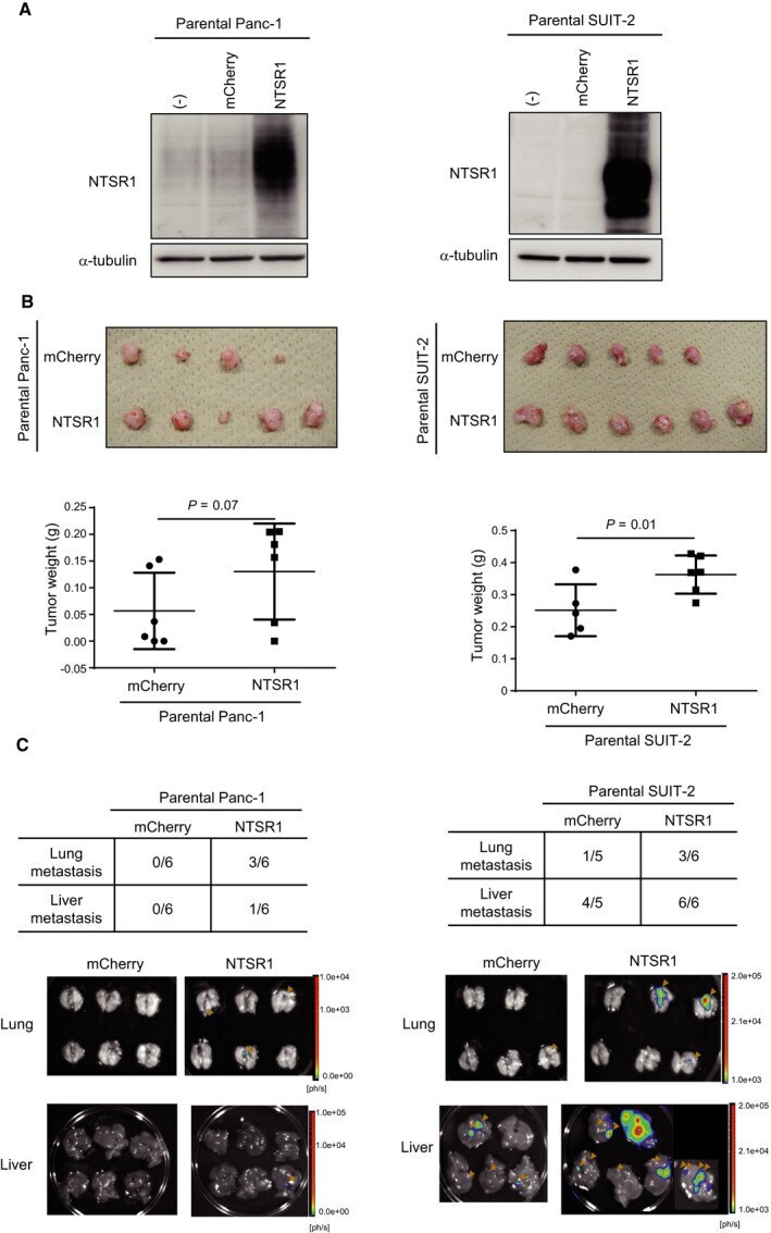
- Fig. 3 Overexpression of NTSR1 promotes tumorigenicity and metastasis of pancreatic cancer cells. (A) The expression of NTSR1 in NTSR1-overexpressing cells. NTSR1 or control mCherry was overexpressed in parental Panc-1 and SUIT-2 cells using a lentiviral vector system. Increased expression of NTSR1 was confirmed by immunoblotting (left: Panc-1, right: SUIT-2). (B) Tumor-forming ability of NTSR1-overexpressing parental Panc-1 or SUIT-2 cells. mCherry/NTSR1-overexpressing parental Panc-1 or SUIT-2 cells were inoculated into the pancreas orthotopically ( n = 5-6 per group). Mice bearing Panc-1-mCherry or Panc-1-NTSR1 cells were euthanized after 2 months. Mice bearing SUIT-2-mCherry or SUIT-2-NTSR1 cells were euthanized after 1 month. The primary tumors were excised (top; no images are shown for mice without tumor formation). Their weights were measured (bottom). One-sided Student's t-test was used to compare two groups. (C) Metastatic ability of NTSR1-overexpressing parental Panc-1 or SUIT-2 cells. Metastatic tumors in the lungs and livers in mice in (B) were detected by ex vivo bioluminescence imaging. The incidence (top) and images (bottom) of lung and liver metastasis are shown. The bioluminescence signals from lung and liver metastatic tumors are indicated with orange arrowheads.