Explore
 Validate
 Learn
Learn Western blot
 Immunocytochemistry
Antibody data
- Antibody Data
- Antigen structure
- References [2]
- Comments [0]
- Validations
- Immunocytochemistry [2]
- Immunohistochemistry [2]
- Other assay [1]
Submit
Validation data
Reference
Comment
Report error
- Product number
- PA5-30116 - Provider product page

- Provider
- Invitrogen Antibodies
- Product name
- PCBP2 Polyclonal Antibody
- Antibody type
- Polyclonal
- Antigen
- Recombinant full-length protein
- Description
- Recommended positive controls: Jurkat, Raji, K562, NCI-H929. Predicted reactivity: Mouse (100%), Rat (97%), Pig (91%), Bovine (93%). Store product as a concentrated solution. Centrifuge briefly prior to opening the vial.
- Reactivity
- Human, Mouse
- Host
- Rabbit
- Isotype
- IgG
- Vial size
- 100 μL
- Concentration
- 1.03 mg/mL
- Storage
- Store at 4°C short term. For long term storage, store at -20°C, avoiding freeze/thaw cycles.
Submitted references Comprehensive profiling of mRNA splicing indicates that GC content signals altered cassette exon inclusion in Ewing sarcoma.
Internal Ribosome Entry Site Dramatically Reduces Transgene Expression in Hematopoietic Cells in a Position-Dependent Manner.
Graham GT, Selvanathan SP, Zöllner SK, Stahl E, Shlien A, Caplen NJ, Üren A, Toretsky JA
NAR cancer 2022 Mar;4(1):zcab052
NAR cancer 2022 Mar;4(1):zcab052
Internal Ribosome Entry Site Dramatically Reduces Transgene Expression in Hematopoietic Cells in a Position-Dependent Manner.
Zheng Q, Zhang X, Yang H, Xie J, Xie Y, Chen J, Yu C, Zhong C
Viruses 2019 Oct 8;11(10)
Viruses 2019 Oct 8;11(10)
No comments: Submit comment
Supportive validation
- Submitted by
- Invitrogen Antibodies (provider)
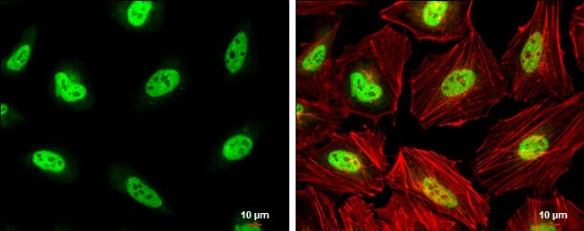
- Main image

- Experimental details
- Immunocytochemistry-Immunofluorescence analysis of PCBP2 was performed in HeLa cells fixed in 4% paraformaldehyde at RT for 15 min. Green: PCBP2 Polyclonal Antibody (Product # PA5-30116) diluted at 1:750. Red: phalloidin, a cytoskeleton marker. Scale bar = 10 µm.
- Submitted by
- Invitrogen Antibodies (provider)
- Main image
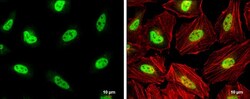
- Experimental details
- Immunocytochemistry-Immunofluorescence analysis of PCBP2 was performed in HeLa cells fixed in 4% paraformaldehyde at RT for 15 min. Green: PCBP2 Polyclonal Antibody (Product # PA5-30116) diluted at 1:750. Red: phalloidin, a cytoskeleton marker. Scale bar = 10 µm.

Supportive validation
- Submitted by
- Invitrogen Antibodies (provider)
- Main image
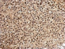
- Experimental details
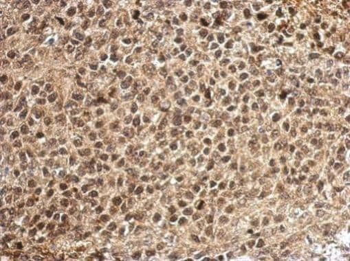
- Immunohistochemical analysis of paraffin-embedded human muscle, using PCBP2 (Product # PA5-30116) antibody at 1:500 dilution. Antigen Retrieval: EDTA based buffer, pH 8.0, 15 min.
- Submitted by
- Invitrogen Antibodies (provider)
- Main image

- Experimental details
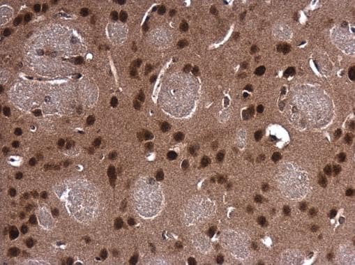
- Immunohistochemistry (Paraffin) analysis of PCBP2 was performed in paraffin-embedded mouse brain tissue using PCBP2 Polyclonal Antibody (Product # PA5-30116) at a dilution of 1:500.
Supportive validation
- Submitted by
- Invitrogen Antibodies (provider)
- Main image

- Experimental details
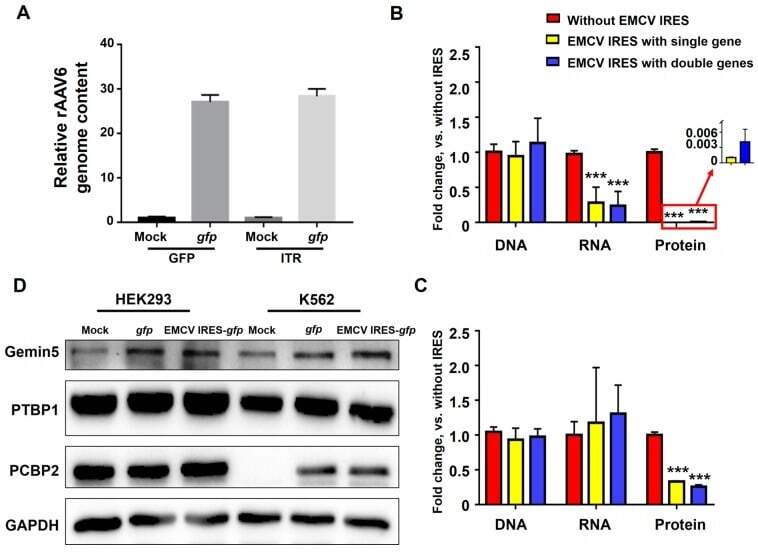
- Figure 5 EMCV IRES inhibited the expression of transgene on the translational level. ( A ) HEK293 cells were transduced with rAAV6-CMVp- gfp at 10,000 vgs/cell. The relative rAAV6 genome content was detected by qPCR using GFP primers and ITR primers. ( B ) K562 and ( C ) HEK293 cells were transduced with rAAV6-CMVp- gfp and rAAV6-CMVp-EMCV IRES- gfp at 10,000 vgs/cell. Total DNA and RNA were isolated at 4 days post-transduction for qPCR. Transgene expression was detected by fluorescence microscopy at 72 hours post-transduction. (D) Western blot of total cell extracts (lysate) from HEK293 cells and K562 cells after rAAV6-CMVp- gfp or rAAV6-CMVp-EMCV IRES- gfp infection for Gemin5, PTBP1, and PCBP2 expression.