Explore
 Validate
 Learn
Learn Western blot
 Immunocytochemistry
Antibody data
- Antibody Data
- Antigen structure
- References [7]
- Comments [0]
- Validations
- Immunocytochemistry [1]
- Immunohistochemistry [2]
Submit
Validation data
Reference
Comment
Report error
- Product number
- HPA007270 - Provider product page

- Provider
- Atlas Antibodies
- Proper citation
- Atlas Antibodies Cat#HPA007270, RRID:AB_1079253
- Product name
- Anti-LSR
- Antibody type
- Polyclonal
- Description
- Polyclonal Antibody against Human LSR, Gene description: lipolysis stimulated lipoprotein receptor, Alternative Gene Names: ILDR3, LISCH7, Validated applications: WB, IHC, ICC, Uniprot ID: Q86X29, Storage: Store at +4°C for short term storage. Long time storage is recommended at -20°C.
- Reactivity
- Human
- Host
- Rabbit
- Conjugate
- Unconjugated
- Isotype
- IgG
- Vial size
- 100 µl
- Concentration
- 0.1 mg/ml
- Storage
- Store at +4°C for short term storage. Long time storage is recommended at -20°C.
- Handling
- The antibody solution should be gently mixed before use.
Submitted references Garcinia cambogia Extract Increased Hepatic Levels of Lipolysis-Stimulated Lipoprotein Receptor and Lipids in Mice on Normal Diet
Macromolecule Translocation across the Intestinal Mucosa of HIV-Infected Patients by Transcytosis and through Apoptotic Leaks.
Lipolysis-Stimulated Lipoprotein Receptor Acts as Sensor to Regulate ApoE Release in Astrocytes
Tricellulin secures the epithelial barrier at tricellular junctions by interacting with actomyosin
Age-related changes in regiospecific expression of Lipolysis Stimulated Receptor (LSR) in mice brain
LSR/angulin-1 is a tricellular tight junction protein involved in blood–brain barrier formation
The role of lipolysis stimulated lipoprotein receptor in breast cancer and directing breast cancer cell behavior.
Hanse M, Akbar S, Layeghkhavidaki H, Yen F
International Journal of Molecular Sciences 2023;24(22):16298
International Journal of Molecular Sciences 2023;24(22):16298
Macromolecule Translocation across the Intestinal Mucosa of HIV-Infected Patients by Transcytosis and through Apoptotic Leaks.
Krug SM, Grünhagen C, Allers K, Bojarski C, Seybold J, Schneider T, Schulzke JD, Epple HJ
Cells 2023 Jul 18;12(14)
Cells 2023 Jul 18;12(14)
Lipolysis-Stimulated Lipoprotein Receptor Acts as Sensor to Regulate ApoE Release in Astrocytes
Herzine A, Sekkat G, Kaminski S, Calcagno G, Boschi-Muller S, Safi H, Corbier C, Siest S, Claudepierre T, Yen F
International Journal of Molecular Sciences 2022;23(15):8630
International Journal of Molecular Sciences 2022;23(15):8630
Tricellulin secures the epithelial barrier at tricellular junctions by interacting with actomyosin
Cho Y, Haraguchi D, Shigetomi K, Matsuzawa K, Uchida S, Ikenouchi J
Journal of Cell Biology 2022;221(4)
Journal of Cell Biology 2022;221(4)
Age-related changes in regiospecific expression of Lipolysis Stimulated Receptor (LSR) in mice brain
Egles C, El Hajj A, Yen F, Oster T, Malaplate C, Pauron L, Corbier C, Lanhers M, Claudepierre T
PLOS ONE 2019;14(6):e0218812
PLOS ONE 2019;14(6):e0218812
LSR/angulin-1 is a tricellular tight junction protein involved in blood–brain barrier formation
Sohet F, Lin C, Munji R, Lee S, Ruderisch N, Soung A, Arnold T, Derugin N, Vexler Z, Yen F, Daneman R
Journal of Cell Biology 2015;208(6):703-711
Journal of Cell Biology 2015;208(6):703-711
The role of lipolysis stimulated lipoprotein receptor in breast cancer and directing breast cancer cell behavior.
Reaves DK, Fagan-Solis KD, Dunphy K, Oliver SD, Scott DW, Fleming JM
PloS one 2014;9(3):e91747
PloS one 2014;9(3):e91747
No comments: Submit comment
Supportive validation
- Submitted by
- Atlas Antibodies (provider)
- Main image
- Experimental details
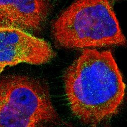
- Immunofluorescent staining of human cell line A-431 shows localization to nucleoplasm, plasma membrane & cytosol.
- Sample type
- Human

Supportive validation
Supportive validation
- Submitted by
- Atlas Antibodies (provider)
- Enhanced method
- Orthogonal validation
- Main image
- Experimental details
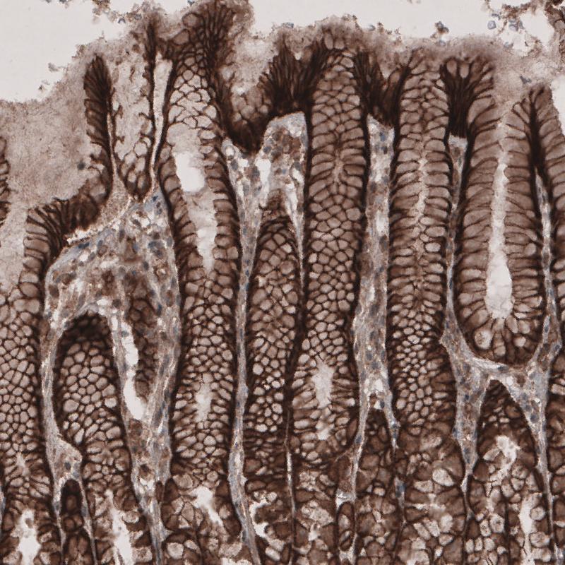
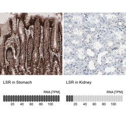
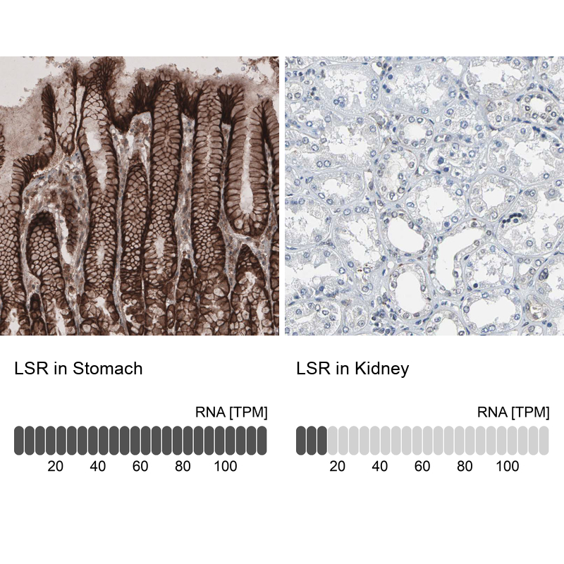
- Immunohistochemistry analysis in human stomach and kidney tissues using HPA007270 antibody. Corresponding LSR RNA-seq data are presented for the same tissues.
- Sample type
- Human
- Protocol
- Protocol
Supportive validation
- Submitted by
- Atlas Antibodies (provider)
- Enhanced method
- Orthogonal validation
- Main image

- Experimental details
- Immunohistochemistry analysis in human stomach and kidney tissues using HPA007270 antibody. Corresponding LSR RNA-seq data are presented for the same tissues.
- Sample type
- Human
- Protocol
- Protocol