Explore
 Validate
 Learn
Learn Western blot
 Immunocytochemistry
Antibody data
- Antibody Data
- Antigen structure
- References [0]
- Comments [0]
- Validations
- Immunocytochemistry [2]
Submit
Validation data
Reference
Comment
Report error
- Product number
- MA5-44745 - Provider product page

- Provider
- Invitrogen Antibodies
- Product name
- PRPF4 Recombinant Rabbit Monoclonal Antibody (JE65-37)
- Antibody type
- Monoclonal
- Antigen
- Synthetic peptide
- Reactivity
- Human
- Host
- Rabbit
- Isotype
- IgG
- Antibody clone number
- JE65-37
- Vial size
- 100 µL
- Concentration
- 1 mg/mL
- Storage
- Store at 4°C short term. For long term storage, store at -20°C, avoiding freeze/thaw cycles.
No comments: Submit comment
Supportive validation
- Submitted by
- Invitrogen Antibodies (provider)
- Main image
- Experimental details
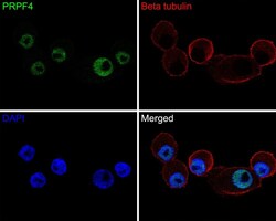
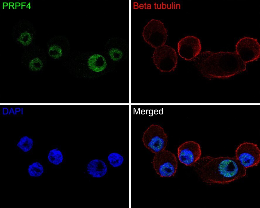
- Immunocytochemistry analysis of PRPF4 in NCI-H1395 cells. Cells were fixed in 4% paraformaldehyde for 10 minutes at 37 ℃, permeabilized with 0.05% Triton X-100 in PBS for 20 minutes, and then blocked with 2% negative goat serum for 30 minutes at room temperature. Samples were incubated in PRPF4 Monoclonal antibody (Product # MA5-44745) using a dilution of 1:50 in 2% negative goat serum overnight at 4 ℃ followed by Goat Anti-Rabbit IgG H&L (Alexa Fluor® 488) secondary antibody at a dilution of 1:1,000. Nuclear DNA was labelled in blue with DAPI.
- Submitted by
- Invitrogen Antibodies (provider)
- Main image
- Experimental details
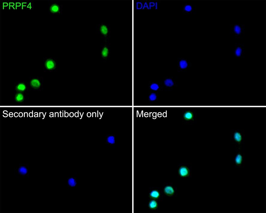
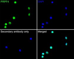
- Immunocytochemistry analysis of PRPF4 in SiHa cells. Cells were fixed in 4% paraformaldehyde for 10 minutes at 37 ℃, permeabilized with 0.05% Triton X-100 in PBS for 20 minutes, and then blocked with 2% negative goat serum for 30 minutes at room temperature. Samples were incubated in PRPF4 Monoclonal antibody (Product # MA5-44745) using a dilution of 1:200 in 2% negative goat serum overnight at 4 ℃ followed by Goat Anti-Rabbit IgG H&L (Alexa Fluor® 488) secondary antibody at a dilution of 1:1,000. PBS instead of the primary antibody was used as the secondary antibody only control. Nuclear DNA was labelled in blue with DAPI.