 Explore
 Validate
 Learn
Learn Western blot
 Flow cytometry
Antibody data
- Antibody Data
- Antigen structure
- References [0]
- Comments [0]
- Validations
- Western blot [1]
- Immunohistochemistry [3]
Submit
Validation data
Reference
Comment
Report error
- Product number
- NBP2-29621 - Provider product page

- Provider
- Novus Biologicals
- Product name
- Mouse Monoclonal BST2 Antibody
- Antibody type
- Monoclonal
- Description
- Protein A or G purified.
- Reactivity
- Human
- Host
- Mouse
- Isotype
- IgG
- Vial size
- 0.1 mg
- Concentration
- 1.0 mg/ml
- Storage
- Store at 4C short term. Aliquot and store at -20C long term. Avoid freeze-thaw cycles.
No comments: Submit comment
Supportive validation
- Submitted by
- Novus Biologicals (provider)
- Main image

- Experimental details
- Western Blot: BST2 Antibody (2E2) [NBP2-29621] - Analysis of BST2 protein in (A) human heart lysate, (B) human ovary lysate and (C) partial recombinant BST2 protein with monoclonal BST2 antibody at a concentration of 1 ug/ml. In the tested human lysates, two distinct bands were observed which represents the glycosylated (lower bands) and the ubiquitinated (higher bands ) forms of BST2 protein.

Supportive validation
- Submitted by
- Novus Biologicals (provider)
- Main image

- Experimental details
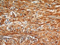
- Immunohistochemistry-Paraffin: BST2 Antibody (2E2) [NBP2-29621] - Analysis of BST2 protein in a section of human lung cancer (squamous cell carcinoma) using 5 ug/ml concentration of BST2 antibody (clone 2E2). The carcinoma cells depicted distinct membrane-cytoplasmic BST2 positivity with more intense staining in the cellular membranes, whereas, mild to moderate staining was observed in tumor stroma also.

- Submitted by
- Novus Biologicals (provider)
- Main image

- Experimental details
- Immunohistochemistry-Paraffin: BST2 Antibody (2E2) [NBP2-29621] - Analysis of BST2 protein in a section of human endometrial carcinoma using 5 ug/ml concentration of BST2 antibody (clone 2E2). The carcinoma cells depicted distinct membrane-cytoplasmic BST2 positivity with more intense staining in the cellular membranes, whereas, weak staining was observed in tumor stroma.

- Submitted by
- Novus Biologicals (provider)
- Main image
- Experimental details
- Immunohistochemistry-Paraffin: BST2 Antibody (2E2) [NBP2-29621] - Analysis of BST2 protein in a section of human renal cell carcinoma (clear cell type) using 5 ug/ml concentration of BST2 antibody (clone 2E2). The carcinoma cells depicted distinct membrane-cytoplasmic BST2 positivity with more intense staining in cellular membranes.
