Explore
 Validate
 Learn
Learn Flow cytometry
 Other assay
Antibody data
- Antibody Data
- Antigen structure
- References [7]
- Comments [0]
- Validations
- Other assay [6]
Submit
Validation data
Reference
Comment
Report error
- Product number
- 16-3179-82 - Provider product page

- Provider
- Invitrogen Antibodies
- Product name
- CD317 (BST2, PDCA-1) Monoclonal Antibody (26F8), Functional Grade, eBioscience™
- Antibody type
- Monoclonal
- Antigen
- Other
- Description
- Description: This 26F8 monoclonal antibody reacts with human CD317 (also known as BST2 and tetherin). CD317 is a 30-36-kDa type II transmembrane protein expressed on B cells and bone marrow stromal cells. Although reports have indicated that CD317 mRNA is detectable in activated T cells, protein expression in primary T cells and macrophages is undetectable. Moreover, certain T cell lines, such as Jurkat, do not express detectable levels of CD317 protein. CD317 has been associated with pre-B cell growth and the terminal differentiation of plasma B cells. More recently, this molecule has been reported to prevent HIV-1 virion release from the surface of infected cells, leading to reuptake and degradation of the virus. This activity is inhibited by the HIV-1 accessory protein Vpu. CD317 has been identified as the ligand for the ILT7 receptor. Applications Reported: This 26F8 antibody has been reported for use in flow cytometric analysis and functional assays. The 26F8 clone has been reported to block the binding of CD317 to the ILT7 receptor. Applications Tested: This 26F8 antibody has been tested by flow cytometric analysis on MCF7 cells. This can be used at less than or equal to 0.125 µg per test. A test is defined as the amount (µg) of antibody that will stain a cell sample in a final volume of 100 µL. Cell number should be determined empirically but can range from 10^5 to 10^8 cells/test. It is recommended that the antibody be carefully titrated for optimal performance in the assay of interest. Storage and handling: Use in a sterile environment. Filtration: 0.2 µm post-manufacturing filtered. Purity: Greater than 90%, as determined by SDS-PAGE. Endotoxin Level: Less than 0.001 ng/µg antibody, as determined by LAL assay. Aggregation: Less than 10%, as determined by HPLC.
- Reactivity
- Human
- Host
- Mouse
- Isotype
- IgG
- Antibody clone number
- 26F8
- Vial size
- 100 µg
- Concentration
- 1 mg/mL
- Storage
- 4° C
Submitted references Japanese encephalitis virus counteracts BST2 restriction via its envelope protein E.
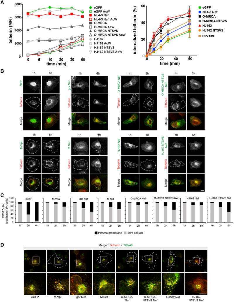
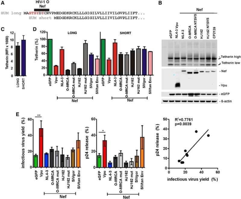
Nef proteins of epidemic HIV-1 group O strains antagonize human tetherin.
Ig-like transcript 7, but not bone marrow stromal cell antigen 2 (also known as HM1.24, tetherin, or CD317), modulates plasmacytoid dendritic cell function in primary human blood leukocytes.
Vpu enhances HIV-1 virus release in the absence of Bst-2 cell surface down-modulation and intracellular depletion.
Tetherin inhibits retrovirus release and is antagonized by HIV-1 Vpu.
The interferon-induced protein BST-2 restricts HIV-1 release and is downregulated from the cell surface by the viral Vpu protein.
Molecular cloning and chromosomal mapping of a bone marrow stromal cell surface gene, BST2, that may be involved in pre-B-cell growth.
Li M, Wang P, Zheng Z, Hu K, Zhang M, Guan X, Fu M, Zhang D, Wang W, Xiao G, Hu Q, Liu Y
Virology 2017 Oct;510:67-75
Virology 2017 Oct;510:67-75
Nef proteins of epidemic HIV-1 group O strains antagonize human tetherin.
Kluge SF, Mack K, Iyer SS, Pujol FM, Heigele A, Learn GH, Usmani SM, Sauter D, Joas S, Hotter D, Bibollet-Ruche F, Plenderleith LJ, Peeters M, Geyer M, Sharp PM, Fackler OT, Hahn BH, Kirchhoff F
Cell host & microbe 2014 Nov 12;16(5):639-50
Cell host & microbe 2014 Nov 12;16(5):639-50
Ig-like transcript 7, but not bone marrow stromal cell antigen 2 (also known as HM1.24, tetherin, or CD317), modulates plasmacytoid dendritic cell function in primary human blood leukocytes.
Tavano B, Galao RP, Graham DR, Neil SJ, Aquino VN, Fuchs D, Boasso A
Journal of immunology (Baltimore, Md. : 1950) 2013 Mar 15;190(6):2622-30
Journal of immunology (Baltimore, Md. : 1950) 2013 Mar 15;190(6):2622-30
Vpu enhances HIV-1 virus release in the absence of Bst-2 cell surface down-modulation and intracellular depletion.
Miyagi E, Andrew AJ, Kao S, Strebel K
Proceedings of the National Academy of Sciences of the United States of America 2009 Feb 24;106(8):2868-73
Proceedings of the National Academy of Sciences of the United States of America 2009 Feb 24;106(8):2868-73
Tetherin inhibits retrovirus release and is antagonized by HIV-1 Vpu.
Neil SJ, Zang T, Bieniasz PD
Nature 2008 Jan 24;451(7177):425-30
Nature 2008 Jan 24;451(7177):425-30
The interferon-induced protein BST-2 restricts HIV-1 release and is downregulated from the cell surface by the viral Vpu protein.
Van Damme N, Goff D, Katsura C, Jorgenson RL, Mitchell R, Johnson MC, Stephens EB, Guatelli J
Cell host & microbe 2008 Apr 17;3(4):245-52
Cell host & microbe 2008 Apr 17;3(4):245-52
Molecular cloning and chromosomal mapping of a bone marrow stromal cell surface gene, BST2, that may be involved in pre-B-cell growth.
Ishikawa J, Kaisho T, Tomizawa H, Lee BO, Kobune Y, Inazawa J, Oritani K, Itoh M, Ochi T, Ishihara K
Genomics 1995 Apr 10;26(3):527-34
Genomics 1995 Apr 10;26(3):527-34
No comments: Submit comment
Supportive validation
- Submitted by
- Invitrogen Antibodies (provider)
- Main image
- Experimental details
- NULL
- Submitted by
- Invitrogen Antibodies (provider)
- Main image

- Experimental details
- NULL
- Submitted by
- Invitrogen Antibodies (provider)
- Main image
- Experimental details
- NULL

- Submitted by
- Invitrogen Antibodies (provider)
- Main image

- Experimental details
- NULL
- Submitted by
- Invitrogen Antibodies (provider)
- Main image

- Experimental details
- NULL
- Submitted by
- Invitrogen Antibodies (provider)
- Main image
- Experimental details
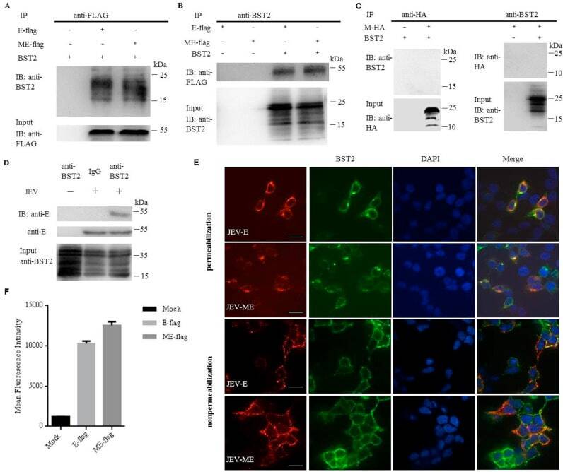
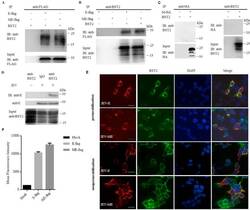
- Fig. 5 JEV protein E physically interacts with BST2. 293T cells were cotransfected with pBST2 and plasmid expressing E-flag, ME-flag or M-HA. At 48 h post transfection, cell lysates were analyzed by co-IP. Co-IP was pulled down using the anti-BST2, anti-flag or anti-HA antibody. Proteins were immunoprecipitated with the anti-flag (A) or anti-BST2 antibody (B) as indicated. (C) Proteins were immunoprecipitated with the anti-HA or anti-BST2 antibody as indicated. (D) HeLa cells were infected with JEV at a MOI of 25. At 48 h post infection, cell lysates were analyzed by co-IP. Co-IP was pulled down using the anti-BST2 antibody. One representative experiment out of three is shown. (E) Colocalization of BST2 with JEV E-flag or ME-flag. 293T cells cotransfected with pBST2 and plasmid expressing E-flag or ME-flag were costained with anti-flag (red) and anti-BST2 (green) antibodies. Nuclei were counterstained with DAPI (blue). Representative confocal images from three independent experiments are shown. Scale bars in all panels represent 10 um. (F) HeLa cells were transfected with plasmid expressing protein E. The surface expression of BST2 was analyzed by flow cytometry. Fig. 5