 Explore
 Validate
 Learn
Learn Western blot
 ELISA
ELISA Immunocytochemistry
Antibody data
- Antibody Data
- Antigen structure
- References [0]
- Comments [0]
- Validations
- Immunocytochemistry [2]
- Immunoprecipitation [1]
- Immunohistochemistry [4]
- Other assay [1]
Submit
Validation data
Reference
Comment
Report error
- Product number
- PA5-80385 - Provider product page

- Provider
- Invitrogen Antibodies
- Product name
- BST-2 Polyclonal Antibody
- Antibody type
- Polyclonal
- Antigen
- Recombinant full-length protein
- Description
- This product is preservative free. It is recommended to add sodium azide to avoid contamination (final concentration 0.05%-0.1%). This antibody has specificity for Human BST2.
- Reactivity
- Human
- Host
- Rabbit
- Isotype
- IgG
- Vial size
- 100 μL
- Concentration
- 1 mg/mL
- Storage
- Store at 4°C short term. For long term storage, store at -20°C, avoiding freeze/thaw cycles.
No comments: Submit comment
Supportive validation
- Submitted by
- Invitrogen Antibodies (provider)
- Main image

- Experimental details
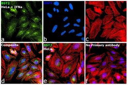
- Immunofluorescence analysis of Bone marrow stromal antigen 2 was performed using 70% confluent log phase HeLa cells treated with 1 ng/mL IFNa for 24 Hrs. The cells were fixed with 4% paraformaldehyde for 10 minutes, permeabilized with 0.1% Triton™ X-100 for 10 minutes, and blocked with 2% BSA for 1 hour at room temperature. The cells were labeled with BST-2 Polyclonal Antibody (Product # PA5-80385) at 1:200 dilution in 0.1% BSA, incubated at 4 degree celsius overnight and then labeled with Donkey anti-Rabbit IgG (H+L) Highly Cross-Adsorbed Secondary Antibody, Alexa Fluor Plus 488 (Product # A32790), (1:2000 dilution), for 45 minutes at room temperature (Panel a: Green). Nuclei (Panel b:Blue) were stained with ProLong™ Diamond Antifade Mountant with DAPI (Product # P36962). F-actin (Panel c: Red) was stained with Rhodamine Phalloidin (Product # R415, 1:300). Panel d represents the merged image showing Nucleus and cytoplasm localization. Panel e represents untreated cells with reduced signal. Panel f represents control cells with no primary antibody to assess background. The images were captured at 60x magnification.

- Submitted by
- Invitrogen Antibodies (provider)
- Main image
- Experimental details
- Immunofluorescence analysis of Bone marrow stromal antigen 2 was performed using 70% confluent log phase HeLa cells treated with 1 ng/mL IFNa for 24 Hrs. The cells were fixed with 4% paraformaldehyde for 10 minutes, permeabilized with 0.1% Triton™ X-100 for 10 minutes, and blocked with 2% BSA for 1 hour at room temperature. The cells were labeled with BST-2 Polyclonal Antibody (Product # PA5-80385) at 1:200 dilution in 0.1% BSA, incubated at 4 degree celsius overnight and then labeled with Donkey anti-Rabbit IgG (H+L) Highly Cross-Adsorbed Secondary Antibody, Alexa Fluor Plus 488 (Product # A32790), (1:2000 dilution), for 45 minutes at room temperature (Panel a: Green). Nuclei (Panel b:Blue) were stained with ProLong™ Diamond Antifade Mountant with DAPI (Product # P36962). F-actin (Panel c: Red) was stained with Rhodamine Phalloidin (Product # R415, 1:300). Panel d represents the merged image showing Nucleus and cytoplasm localization. Panel e represents untreated cells with reduced signal. Panel f represents control cells with no primary antibody to assess background. The images were captured at 60x magnification.

Supportive validation
- Submitted by
- Invitrogen Antibodies (provider)
- Main image

- Experimental details
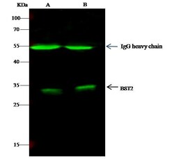
- BST-2 Immunoprecipitation using: Lane A: 0.5 mg Hela Whole Cell Lysate, Lane B: 0.5 mg Jurkat Whole Cell Lysate 1 µL with BST-2 Polyclonal Antibody (Product # PA5-80385) and 60 μg of Immunomagnetic beads Protein G. Primary antibody: BST-2 Polyclonal Antibody, at 1:500 dilution. Secondary antibody: Dylight 800-labeled antibody to rabbit IgG (H+L), at 1:5,000 dilution. Developed using the Odyssey technique. Performed under reducing conditions. Predicted band size: 20 kDa. Observed band size: 28 kDa.

Supportive validation
- Submitted by
- Invitrogen Antibodies (provider)
- Main image
- Experimental details
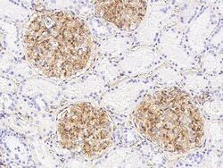
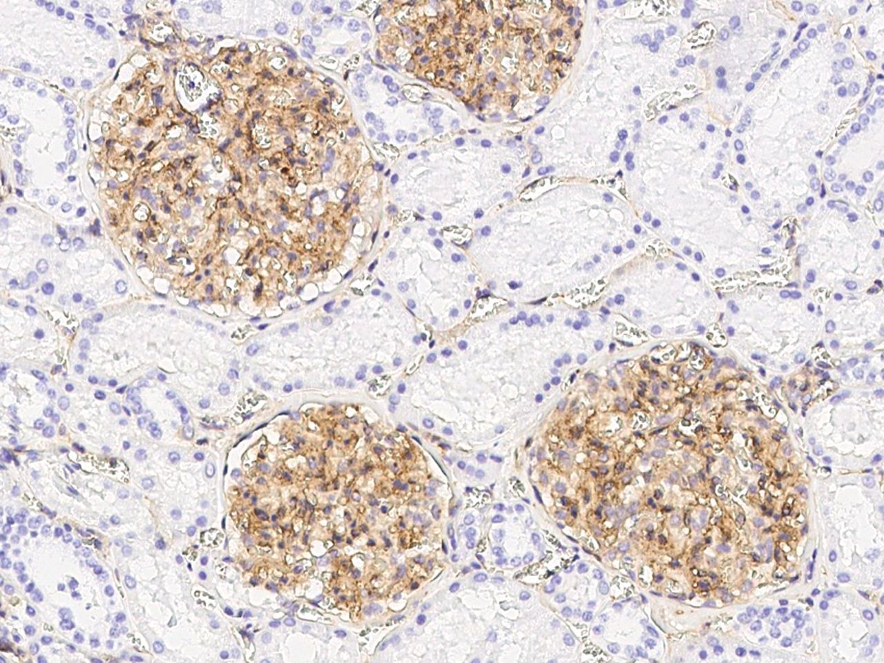
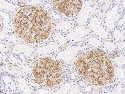
- Immunohistochemical staining of human BST-2 in human kidney with BST-2 Polyclonal Antibody (Product # PA5-80385, 1:5,000 dilution, formalin-fixed paraffin embedded sections).

- Submitted by
- Invitrogen Antibodies (provider)
- Main image
- Experimental details
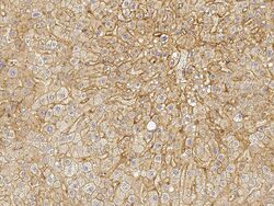
- Immunohistochemical staining of human BST-2 in human liver with BST-2 Polyclonal Antibody (Product # PA5-80385, 1:5,000 dilution, formalin-fixed paraffin embedded sections).
- Submitted by
- Invitrogen Antibodies (provider)
- Main image

- Experimental details
- Immunohistochemical staining of human BST-2 in human kidney with BST-2 Polyclonal Antibody (Product # PA5-80385, 1:5,000 dilution, formalin-fixed paraffin embedded sections).

- Submitted by
- Invitrogen Antibodies (provider)
- Main image
- Experimental details
- Immunohistochemical staining of human BST-2 in human liver with BST-2 Polyclonal Antibody (Product # PA5-80385, 1:5,000 dilution, formalin-fixed paraffin embedded sections).

Supportive validation
- Submitted by
- Invitrogen Antibodies (provider)
- Main image
- Experimental details
- BST-2 Immunoprecipitation using: Lane A: 0.5 mg Hela Whole Cell Lysate, Lane B: 0.5 mg Jurkat Whole Cell Lysate 1 µL with BST-2 Polyclonal Antibody (Product # PA5-80385) and 60 μg of Immunomagnetic beads Protein G. Primary antibody: BST-2 Polyclonal Antibody, at 1:500 dilution. Secondary antibody: Dylight 800-labeled antibody to rabbit IgG (H+L), at 1:5,000 dilution. Developed using the Odyssey technique. Performed under reducing conditions. Predicted band size: 20 kDa. Observed band size: 28 kDa.
