 Explore
 Validate
 Learn
Learn Western blot
 Flow cytometry
Antibody data
- Antibody Data
- Antigen structure
- References [1]
- Comments [0]
- Validations
- Western blot [2]
- Immunohistochemistry [2]
- Other assay [2]
Submit
Validation data
Reference
Comment
Report error
- Product number
- PA5-23505 - Provider product page

- Provider
- Invitrogen Antibodies
- Product name
- BST-2 Polyclonal Antibody
- Antibody type
- Polyclonal
- Antigen
- Other
- Reactivity
- Human, Mouse, Rat
- Host
- Rabbit
- Isotype
- IgG
- Vial size
- 100 μg
- Concentration
- 1 mg/mL
- Storage
- Store at 4°C short term. For long term storage, store at -20°C, avoiding freeze/thaw cycles.
Submitted references BST2 Promotes Tumor Growth via Multiple Pathways in Hepatocellular Carcinoma.
Xu X, Wang Y, Xue F, Guan E, Tian F, Xu J, Zhang H
Cancer investigation 2020 May;38(5):329-337
Cancer investigation 2020 May;38(5):329-337
No comments: Submit comment
Supportive validation
- Submitted by
- Invitrogen Antibodies (provider)
- Main image
- Experimental details
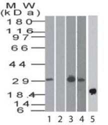
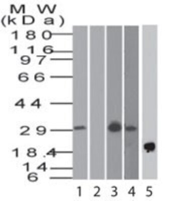
- Western blot analysis of BST-2 in human heart in the 1) absence and 2) presence of immunizing peptide, 3) human Jurkat, 4) mouse RAW lysate and 5) recombinant human BST2 protein. Samples were incubated in BST-2 polyclonal antibody (Product # PA5-23505) using a dilution of 4 µg/mL followed by a Goat anti-rabbit IgG HRP secondary antibody. PicoTect ECL substrate solution was used for this test.
- Submitted by
- Invitrogen Antibodies (provider)
- Main image

- Experimental details
- Western blot analysis of BST-2 (CD317) in 0.5 mg/mL Jurkat lysate. Samples were incubated in BST-2 (CD317) polyclonal antibody (Product # PA5-23505). This experiment was performed under standard reducing conditions using the 12-230 kDa separation system.

Supportive validation
- Submitted by
- Invitrogen Antibodies (provider)
- Main image

- Experimental details
- Immunohistochemical analysis of BST-2 in human liver tissue. Samples were incubated in BST-2 polyclonal antibody (Product # PA5-23505) using a dilution of 5 µg/mL.

- Submitted by
- Invitrogen Antibodies (provider)
- Main image

- Experimental details
- Immunohistochemical analysis of BST-2 in human liver tissue. Samples were incubated in BST-2 polyclonal antibody (Product # PA5-23505) using a dilution of 5 µg/mL.

Supportive validation
- Submitted by
- Invitrogen Antibodies (provider)
- Main image
- Experimental details
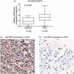
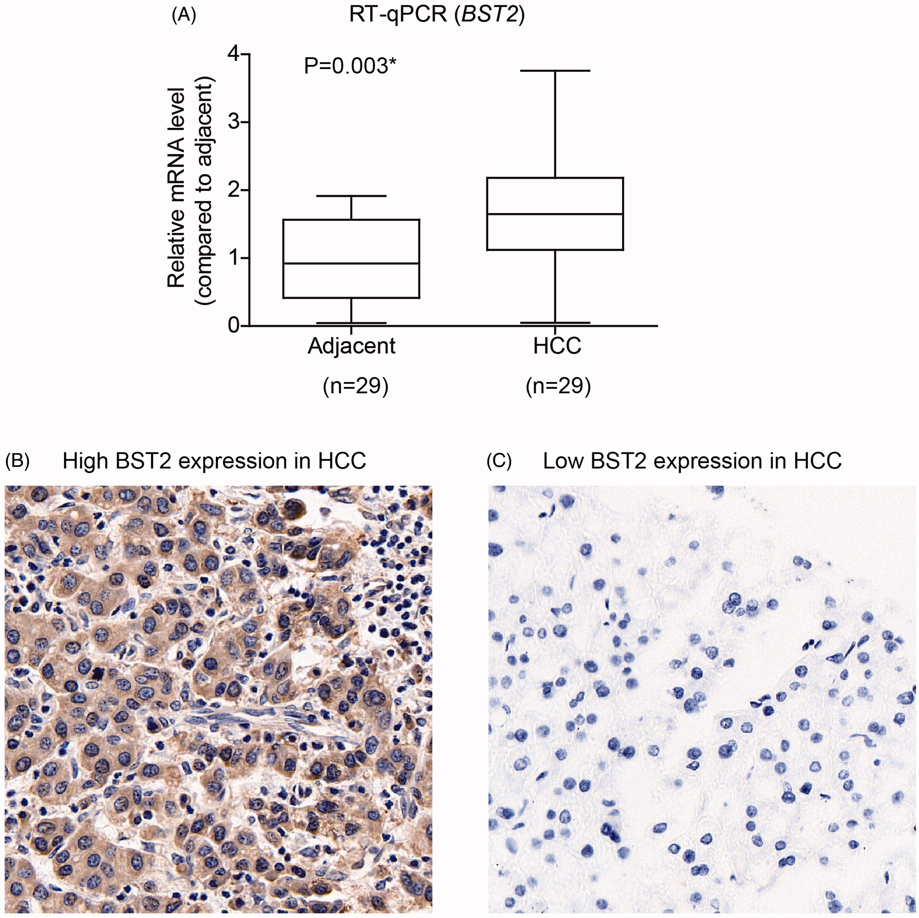
- Figure 1. Analyses of BST2 expression in patients with HCC. (A) Total mRNA level of BST2 were examined in HCC tissues together with adjacent normal liver tissues by real-time qPCR. (B, C) IHC staining results showed representative high and low expression of BST2 in HCC tissues. Magnification, 400x. * p < 0.001 by Student's t -test.
- Submitted by
- Invitrogen Antibodies (provider)
- Main image
- Experimental details
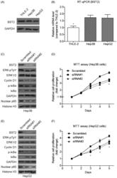
- Figure 3. BST2 promotes proliferation capacity of HCC cell line. (A, B) The protein levels and mRNA levels of BST2 in HCC cell lines and control cell line were measured by Western blotting and real-time qPCR. (C,E) Western Blot results showed the protein levels of BST2, pERK, cyclin D1, p- IkBa, total IkBa, and p65-NF-kB in BST2 knock down- cells. (D, F) Cell proliferation ability was tested in BST2 knockdown cells.
