 Explore
 Validate
 Learn
Learn ELISA
ELISA Other assay
Antibody data
- Antibody Data
- Antigen structure
- References [1]
- Comments [0]
- Validations
- Other assay [3]
Submit
Validation data
Reference
Comment
Report error
- Product number
- PA1-18312 - Provider product page

- Provider
- Invitrogen Antibodies
- Product name
- Sortilin Polyclonal Antibody
- Antibody type
- Polyclonal
- Antigen
- Other
- Description
- Reconstitute in 100 µL of sterile water. Centrifuge to remove any insoluble material. After reconstitution keep aliquots at -20 °C for a higher stability, and at 4 °C with an appropriate antibacterial agent. Glycerol (1:1) may be added for an additional stability. Avoid repetitive freeze/thaw cycles.
- Reactivity
- Human, Mouse, Rat
- Host
- Rabbit
- Isotype
- IgG
- Vial size
- 100 µL
- Concentration
- Conc. Not Determined
- Storage
- -20° C, Avoid Freeze/Thaw Cycles
Submitted references Secreted gelsolin desensitizes and induces apoptosis of infiltrated lymphocytes in prostate cancer.
Chen CC, Chiou SH, Yang CL, Chow KC, Lin TY, Chang HW, You WC, Huang HW, Chen CM, Chen NC, Chou FP, Chou MC
Oncotarget 2017 Sep 29;8(44):77152-77167
Oncotarget 2017 Sep 29;8(44):77152-77167
No comments: Submit comment
Supportive validation
- Submitted by
- Invitrogen Antibodies (provider)
- Main image

- Experimental details
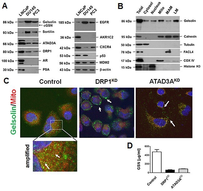
- Figure 2 Gelsolin expression in PCa cell lines, and the intracellular locations and extracellular levels of gelsolin (A) Expression of gelsolin and tumor-associated proteins in three PCa cell lines, LNCaP, DU145 and PC3, were determined by Western blotting. Gelsolin expression correlated positively with sortilin and EGFR expression, but negatively with ATAD3A and DRP1 expression (essential proteins for intracellular transport [ 7 ]). (B) Percoll self-generating gradient fractionation was used to localize gelsolin in the fractions of cytosol, nucleus, mitochondria-associated membrane (MAM), mitochondria, and light membranes (LM, majority is microsomes), suggesting that most of gelsolin was present in membrane structures. (C) Confocal immunofluorescence micrographs revealed that gelsolin was localized in vesicles, and some of gelsolin signal overlapped with the mitochondrial marker (Control, the left panel and the amplified region). Nuclei were stained with 4',6-diamidino-2-phenylindole (DAPI). Knockdown of DRP1 (DRP1 KD ) increased the numbers of vacuoles, suggesting that the ER or MAM were enlarged (center panel). When ATAD3A was knocked down (ATAD3A KD ), mitochondrial staining was reduced and the gelsolin signals were randomly distributed in the cytoplasmic vesicles (right panel). (D) Silencing of ATAD3A or DRP1 markedly reduced extracellular gelsolin levels in culture media of LNCaP cells. The results are shown as the means +-standard deviations of three independent e
- Submitted by
- Invitrogen Antibodies (provider)
- Main image

- Experimental details
- Figure 6 Intracellularly, sortilin binds to WASF3 and ATAD3A to promote the intake of cholesterol (A) Antibodies specific to sortilin, were used to co-precipitate ATAD3A and WASF3 from the cytoplasmic proteins of DU145 cells. (B) Various deletion mutants were constructed for ATAD3A and WASF3, which were respectively tagged with c - myc and human influenza hemagglutinin (HA), and the interactions between these proteins were examined. (C) Only full-length WASF3 co-precipitated with ATAD3A and sortilin (upper panel). ATAD3A did not co-precipitate with the deletion mutants DWN150 (without N-terminal 150 amino acid residues) and DWN220 of WASF3, although sortilin still co-precipitated with DWN150. The lower panel demonstrates the protein levels of the WASF3 mutants. (D) Wild-type ATAD3A co-precipitated with WASF3 and sortilin (upper panel). The deletion mutant DAN220 lost the ability to co-precipitate with WASF3 but not sortilin. The lower panel depicts the protein levels of ATAD3A. (E) Filipin III (FR4767, Sigma) fluorescent staining of cholesterol and lipoproteins. In control cells, filipin III fluorescence was detected from the plasma membrane to the nuclear envelope. The addition of secreted gelsolin (sGSN) increased the total amount of filipin III fluorescence, indicating that gelsolin increased the intake of cholesterol and lipoproteins. Knockdown of sortilin (SORT KD ), WASF-3 or ATAD3A reduced filipin III fluorescence. In WASF3 KD cells, though some filipin III staining wa

- Submitted by
- Invitrogen Antibodies (provider)
- Main image

- Experimental details
- Figure 7 The effect of AF38469, a SMI of sortilin, on the expression of gelsolin, sortilin and ATAD3A (A) Gelsolin expression was inhibited in a dose-dependent manner by AF38469. (B) AF38469 inhibited gelsolin expression in a time-dependent manner, and also suppressed the expression of ATAD3A and sortilin. The inhibition of ATAD3A expression occurred prior to that of gelsolin and sortilin. (C) AF38469 induced PCa cell death, with a median cytotoxicity 2.5 muM for LNCaP, 1.0 muM for DU145 and 0.82 muM for PC3 cells. AF38469 also induced ( (D ) autophagy (as determined by an increase of acridine orange staining) and ( (E ) nuclear translocation of AIF, a molecular signature of apoptosis (as assessed by an increase of nuclear immunocytochemical staining). (F) Silencing of ATAD3A clearly reduced gelsolin expression, but only marginally affected sortilin. (G) Silencing of sortilin, on the other hand, reduced gelsolin expression, but did not affect that of ATAD3A. Supplementary Materials for ""Secreted gelsolin desensitizes and induces apoptosis of the infiltrated lymphocytes in the prostate cancer""
