 Explore
 Validate
 Learn
Learn Western blot
Antibody data
- Antibody Data
- Antigen structure
- References [1]
- Comments [0]
- Validations
- Western blot [4]
Submit
Validation data
Reference
Comment
Report error
- Product number
- MAB3154 - Provider product page

- Provider
- R&D Systems
- Product name
- Human/Mouse/Rat Sortilin Antibody
- Antibody type
- Monoclonal
- Description
- Protein A or G purified from hybridoma culture supernatant. Detects human, mouse, and rat Sortilin in Western blots.
- Reactivity
- Human, Mouse, Rat
- Host
- Mouse
- Conjugate
- Unconjugated
- Antigen sequence
Q99523- Isotype
- IgG
- Antibody clone number
- 334708
- Vial size
- 100 ug
- Concentration
- LYOPH
- Storage
- Use a manual defrost freezer and avoid repeated freeze-thaw cycles. 12 months from date of receipt, -20 to -70 °C as supplied. 1 month, 2 to 8 °C under sterile conditions after reconstitution. 6 months, -20 to -70 °C under sterile conditions after reconstitution.
Submitted references A Proteomic Survey Indicates Sortilin as a Secondary Substrate of the ER Translocation Inhibitor Cyclotriazadisulfonamide (CADA).
Van Puyenbroeck V, Claeys E, Schols D, Bell TW, Vermeire K
Molecular & cellular proteomics : MCP 2017 Feb;16(2):157-167
Molecular & cellular proteomics : MCP 2017 Feb;16(2):157-167
No comments: Submit comment
Supportive validation
- Submitted by
- R&D Systems (provider)
- Main image
- Experimental details
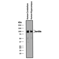
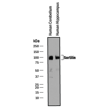
- Detection of Human Sortilin by Western Blot. Western blot shows lysates of human brain (cerebellum) tissue and human brain (hippocampus) tissue. PVDF membrane was probed with 0.25 µg/mL of Mouse Anti-Human/Mouse/Rat Sortilin Monoclonal Antibody (Catalog # MAB3154) followed by HRP-conjugated Anti-Mouse IgG Secondary Antibody (Catalog # HAF018). A specific band was detected for Sortilin at approximately 95-105 kDa (as indicated). This experiment was conducted under reducing conditions and using Immunoblot Buffer Group 1.
- Submitted by
- R&D Systems (provider)
- Main image

- Experimental details
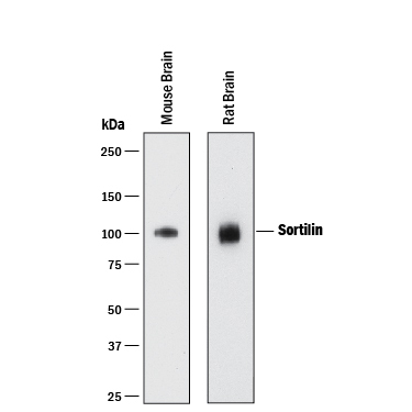
- Detection of Mouse and Rat Sortilin by Western Blot. Western blot shows lysates of mouse brain tissue and rat brain tissue. PVDF membrane was probed with 1 µg/mL of Mouse Anti-Human/Mouse/Rat Sortilin Monoclonal Antibody (Catalog # MAB3154) followed by HRP-conjugated Anti-Mouse IgG Secondary Antibody (Catalog # HAF018). A specific band was detected for Sortilin at approximately 100 kDa (as indicated). This experiment was conducted under reducing conditions and using Immunoblot Buffer Group 1.
- Submitted by
- R&D Systems (provider)
- Main image
- Experimental details
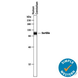
- Detection of Human Sortilin by Simple WesternTM. Simple Western lane view shows lysates of human brain (cerebellum) tissue, loaded at 0.2 mg/mL. A specific band was detected for Sortilin at approximately 87 kDa (as indicated) using 10 µg/mL of Mouse Anti-Human/Mouse/Rat Sortilin Monoclonal Antibody (Catalog # MAB3154) . This experiment was conducted under reducing conditions and using the 12-230 kDa separation system.

- Submitted by
- R&D Systems (provider)
- Main image
- Experimental details
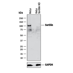
- Western Blot Shows Human Sortilin Specificity by Using Knockout Cell Line. Western blot shows lysates of HeLa human cervical epithelial carcinoma parental cell line and Sortilin knockout HeLa cell line (KO). PVDF membrane was probed with 2 µg/mL of Mouse Anti-Human/Mouse/Rat Sortilin Monoclonal Antibody (Catalog # MAB3154) followed by HRP-conjugated Anti-Mouse IgG Secondary Antibody (Catalog # HAF018). A specific band was detected for Sortilin at approximately 100 kDa (as indicated) in the parental HeLa cell line, but is not detectable in knockout HeLa cell line. GAPDH (Catalog # MAB5718) is shown as a loading control. This experiment was conducted under reducing conditions and using Immunoblot Buffer Group 1.
