Explore
 Validate
 Learn
Learn Western blot
Antibody data
- Antibody Data
- Antigen structure
- References [0]
- Comments [0]
- Validations
- Western blot [2]
- Immunohistochemistry [1]
- Flow cytometry [1]
Submit
Validation data
Reference
Comment
Report error
- Product number
- AGR-052-200UL - Provider product page

- Provider
- Invitrogen Antibodies
- Product name
- GPR84 (extracellular) Polyclonal Antibody
- Antibody type
- Polyclonal
- Antigen
- Other
- Reactivity
- Human, Mouse, Rat
- Host
- Rabbit
- Isotype
- IgG
- Vial size
- 200 µL
- Concentration
- 0.8 mg/mL
- Storage
- -20° C, Avoid Freeze/Thaw Cycles
No comments: Submit comment
Supportive validation
- Submitted by
- Invitrogen Antibodies (provider)
- Main image
- Experimental details
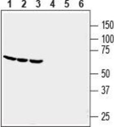
- Western blot analysis of human THP-1 monocytic leukemia (lanes 1 and 4), human HL-60 promyelocytic leukemia (lanes 2 and 5) and human U-87 MG glioblastoma (lanes 3 and 6)cell line lysates: - 1-3. Anti-GPR84 (extracellular) Antibody (#AGR-052), (1:200).4-6. Anti-GPR84 (extracellular) Antibody , preincubated with GPR84 (extracellular) Blocking Peptide (#BLP-GR052).

- Submitted by
- Invitrogen Antibodies (provider)
- Main image
- Experimental details
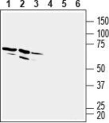
- Western blot analysis of rat brain membranes (lanes 1 and 4), mouse brain membranes (lanes 2 and 5) and rat spleen membranes (lanes 3 and 6): - 1-3. Anti-GPR84 (extracellular) Antibody (#AGR-052), (1:200).4-6. Anti-GPR84 (extracellular) Antibody , preincubated with GPR84 (extracellular) Blocking Peptide (#BLP-GR052).

Supportive validation
- Submitted by
- Invitrogen Antibodies (provider)
- Main image
- Experimental details
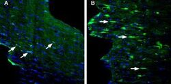
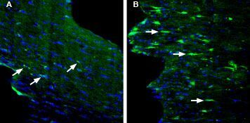
- Expression of GPR84 in mouse fornix in a kainic acid-induced model of temporal lobe epilepsy - Immunohistochemical staining of perfusion-fixed frozen mouse brain sections with Anti-GPR84 (extracellular) Antibody (#AGR-052), (1:200), followed by goat Anti-rabbit-AlexaFluor-488. A. In untreated mouse sections GPR84 immunoreactivity (green) is detected in astrocyte profiles (arrows). B. GPR84 staining (green) in mouse sections obtained four days post kainate-induced seizures, shows an increase in number and size of astrocyte profiles stained for GPR84 (arrows). Cell nuclei are stained with DAPI (blue).
Supportive validation
- Submitted by
- Invitrogen Antibodies (provider)
- Main image

- Experimental details
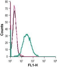
- Cell surface detection of GPR84 in live intact mouse J774 macrophage cells: - (black line) cells. (red) Cells + goat- Anti-rabbit-FITC. (green) Cells + Anti-GPR84 (extracellular) Antibody (#AGR-052), (2.5 µg) + goat- Anti-rabbit-FITC.