Explore
 Validate
 Learn
Learn Western blot
 Immunocytochemistry
 Flow cytometry
Antibody data
- Antibody Data
- Antigen structure
- References [7]
- Comments [0]
- Validations
- Immunocytochemistry [1]
- Other assay [2]
Submit
Validation data
Reference
Comment
Report error
- Product number
- MA1-06315 - Provider product page

- Provider
- Invitrogen Antibodies
- Product name
- Cytokeratin 7 Monoclonal Antibody (RCK105)
- Antibody type
- Monoclonal
- Antigen
- Other
- Description
- MA1-06315 detects cytokeratin 7 in human, mouse, rat, hamster, canine, feline, goat and zebrafish samples. MA1-06315 has sucessfully been used in Western blotting, flow cytometry, immunocytochemistry and immunohistochemistry. The MA1-06315 immunogens are cytokeratins from the human bladder carcinoma cell line T24. Store at 4ºC or in small aliquots at -20ºC.
- Reactivity
- Human, Mouse, Rat, Canine, Feline, Goat, Hamster, Zebrafish
- Host
- Mouse
- Isotype
- IgG
- Antibody clone number
- RCK105
- Vial size
- 100 µL
- Concentration
- 1 mg/mL
- Storage
- Store at 4°C short term. For long term storage, store at -20°C, avoiding freeze/thaw cycles.
Submitted references Differences in Glycolysis and Mitochondrial Respiration between Cytotrophoblast and Syncytiotrophoblast In-Vitro: Evidence for Sexual Dimorphism.
The intact parasympathetic nerve promotes submandibular gland regeneration through ductal cell proliferation.
p16ink4 and cytokeratin 7 immunostaining in predicting HSIL outcome for low-grade squamous intraepithelial lesions: a case series, literature review and commentary.
Microanatomy of the cervical and anorectal squamocolumnar junctions: a proposed model for anatomical differences in HPV-related cancer risk.
Understanding the role of keratins 8 and 18 in neoplastic potential of breast cancer derived cell lines.
A novel blueprint for 'top down' differentiation defines the cervical squamocolumnar junction during development, reproductive life, and neoplasia.
A discrete population of squamocolumnar junction cells implicated in the pathogenesis of cervical cancer.
Bucher M, Kadam L, Ahuna K, Myatt L
International journal of molecular sciences 2021 Oct 8;22(19)
International journal of molecular sciences 2021 Oct 8;22(19)
The intact parasympathetic nerve promotes submandibular gland regeneration through ductal cell proliferation.
Wang X, Li Z, Shao Q, Zhang C, Wang J, Han Z, Wang S, Qin L
Cell proliferation 2021 Jul;54(7):e13078
Cell proliferation 2021 Jul;54(7):e13078
p16ink4 and cytokeratin 7 immunostaining in predicting HSIL outcome for low-grade squamous intraepithelial lesions: a case series, literature review and commentary.
Huang EC, Tomic MM, Hanamornroongruang S, Meserve EE, Herfs M, Crum CP
Modern pathology : an official journal of the United States and Canadian Academy of Pathology, Inc 2016 Dec;29(12):1501-1510
Modern pathology : an official journal of the United States and Canadian Academy of Pathology, Inc 2016 Dec;29(12):1501-1510
Microanatomy of the cervical and anorectal squamocolumnar junctions: a proposed model for anatomical differences in HPV-related cancer risk.
Yang EJ, Quick MC, Hanamornroongruang S, Lai K, Doyle LA, McKeon FD, Xian W, Crum CP, Herfs M
Modern pathology : an official journal of the United States and Canadian Academy of Pathology, Inc 2015 Jul;28(7):994-1000
Modern pathology : an official journal of the United States and Canadian Academy of Pathology, Inc 2015 Jul;28(7):994-1000
Understanding the role of keratins 8 and 18 in neoplastic potential of breast cancer derived cell lines.
Iyer SV, Dange PP, Alam H, Sawant SS, Ingle AD, Borges AM, Shirsat NV, Dalal SN, Vaidya MM
PloS one 2013;8(1):e53532
PloS one 2013;8(1):e53532
A novel blueprint for 'top down' differentiation defines the cervical squamocolumnar junction during development, reproductive life, and neoplasia.
Herfs M, Vargas SO, Yamamoto Y, Howitt BE, Nucci MR, Hornick JL, McKeon FD, Xian W, Crum CP
The Journal of pathology 2013 Feb;229(3):460-8
The Journal of pathology 2013 Feb;229(3):460-8
A discrete population of squamocolumnar junction cells implicated in the pathogenesis of cervical cancer.
Herfs M, Yamamoto Y, Laury A, Wang X, Nucci MR, McLaughlin-Drubin ME, Münger K, Feldman S, McKeon FD, Xian W, Crum CP
Proceedings of the National Academy of Sciences of the United States of America 2012 Jun 26;109(26):10516-21
Proceedings of the National Academy of Sciences of the United States of America 2012 Jun 26;109(26):10516-21
No comments: Submit comment
Supportive validation
- Submitted by
- Invitrogen Antibodies (provider)
- Main image
- Experimental details
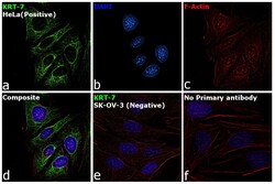
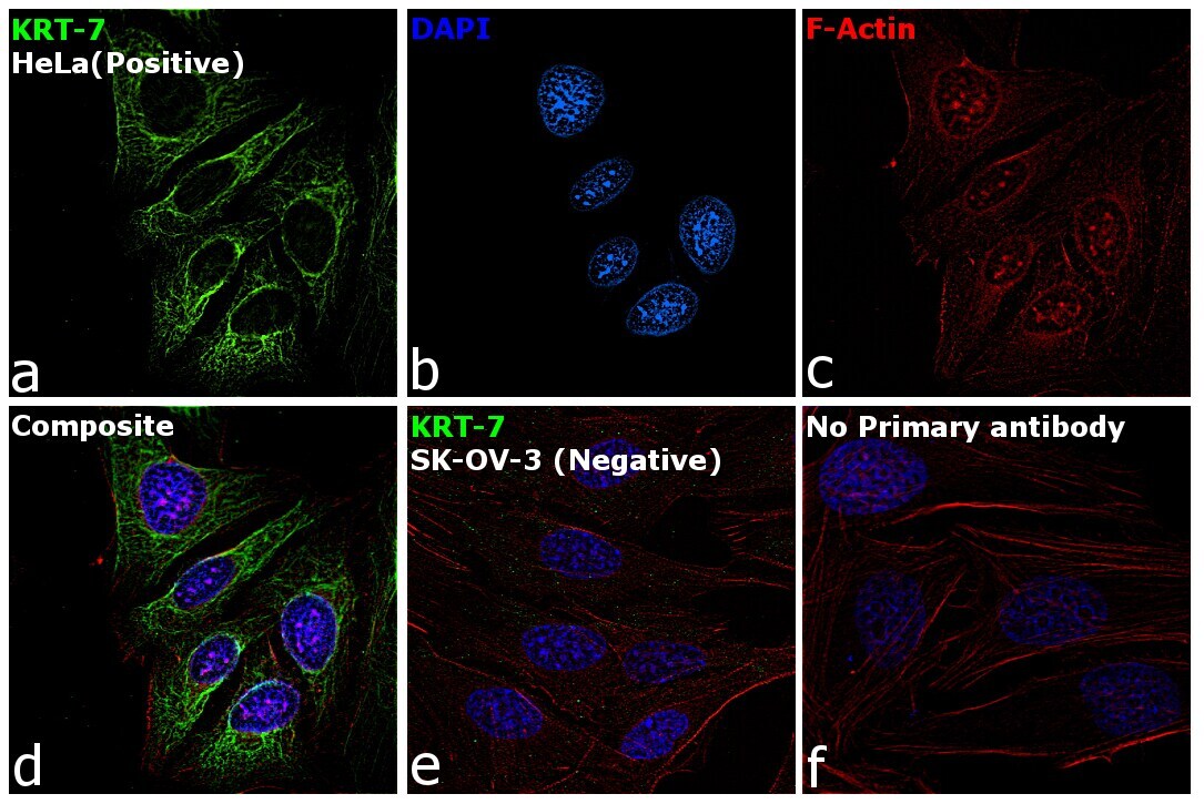
- Immunofluorescence analysis of Cytokeratin 7 was performed using 70% confluent log phase HeLa cells. The cells were fixed with 4% paraformaldehyde for 10 minutes, permeabilized with 0.1% Triton™ X-100 for 15 minutes, and blocked with 2% BSA for 45 minutes at room temperature. The cells were labeled with Cytokeratin 7 Monoclonal Antibody (RCK105) (Product # MA1-06315) at 1:200 dilution in 0.1% BSA, incubated at 4 degree celsius overnight and then labeled with Goat anti-Mouse IgG (H+L) Highly Cross-Adsorbed Secondary Antibody, Alexa Fluor Plus 488 (Product # A32723), (1:2000), for 45 minutes at room temperature (Panel a: Green). Nuclei (Panel b:Red) were stained with ProLong™ Diamond Antifade Mountant with DAPI (Product # P36962). F-actin (Panel c: Blue) was stained with Rhodamine Phalloidin (Product # R415, 1:300). Panel d represents the merged image showing cytoplasmic localization. Panel e represents control cells with no primary antibody to assess background. The images were captured at 60x magnification.
Supportive validation
- Submitted by
- Invitrogen Antibodies (provider)
- Main image
- Experimental details
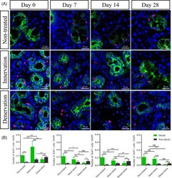
- FIGURE 4 Parasympathetic innervation increases ductal epithelial cell proliferation. (A) Immunofluorescence staining of cytokeratin 7 (green), Ki67 (red) and DAPI (blue) in the submandibular glands during gland regeneration in different groups. Scale bar =20 mum. (B) The number of Ki67+ in ductal and non-ductal cells was different. The relative expression of ki67+ was maintained a stable state at days 14 and 28. *** indicates significance at P < .001, ** P < .01, * P < .05, ns for no significant. Data were shown as mean +- standard deviation

- Submitted by
- Invitrogen Antibodies (provider)
- Main image
- Experimental details
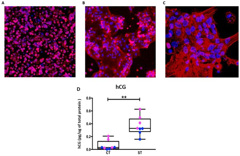
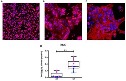
- Figure 1 Identification of trophoblast cells and their syncytialization. ( A ) Cytotrophoblast at 24 h (20x), ( B ) Syncytiotrophoblast at 96 hrs (20x), and ( C ) Syncytiotrophoblast (63x) stained with cytokeratin 7 (red) and counterstained with Hoechst 33,342 for nuclei (blue). ( D ) Human Chorionic Gonadotropin (hCG) production pg of hormone per mug of cell protein. Data presented as minimum, maximum, median, 25th and 75th quartiles boxes, and whisker plots, n = 8, male = blue, female = pink. ** p < 0.01, (Wilcoxon test CT vs. ST).