Explore
 Validate
 Learn
Learn Western blot
 Immunocytochemistry
Antibody data
- Antibody Data
- Antigen structure
- References [0]
- Comments [0]
- Validations
- Western blot [1]
- Immunohistochemistry [2]
- Flow cytometry [3]
Submit
Validation data
Reference
Comment
Report error
- Product number
- NB500-352 - Provider product page

- Provider
- Novus Biologicals
- Proper citation
- Novus Cat#NB500-352, RRID:AB_10001529
- Product name
- Mouse Monoclonal Cytokeratin 7/17 Antibody
- Antibody type
- Monoclonal
- Description
- Protein A purified. The antibody C-46 reacts with Cytokeratin peptides 7 and 17 (54 and 46 kDa). Cytokeratins are a member of intermediate filaments subfamily represented in epithelial tissues.
- Reactivity
- Human, Mouse, Rat, Bovine, Porcine, Rabbit
- Host
- Mouse
- Isotype
- IgG
- Vial size
- 0.1 mg
- Concentration
- 1.0 mg/ml
- Storage
- Store at 4C. Do not freeze.
No comments: Submit comment
Supportive validation
- Submitted by
- Novus Biologicals (provider)
- Main image

- Experimental details
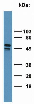
- Western Blot: Cytokeratin 7/17 Antibody (C-46) [NB500-352] - Detection of Cytokeratin 7+17 in HeLa cell lysate with monoclonal antibody C-46.
Supportive validation
- Submitted by
- Novus Biologicals (provider)
- Main image

- Experimental details
- Immunohistochemistry: Cytokeratin 7/17 Antibody (C-46) [NB500-352] - Fig. 1. Immunohistochemistry staining of pancreas (paraffin-embedded sections) with anti-Cytokeratin 7/17 Antibody (C-46).

- Submitted by
- Novus Biologicals (provider)
- Main image

- Experimental details
- Immunohistochemistry-Paraffin: Cytokeratin 7/17 Antibody (C-46) [NB500-352] - Staining of human breast (paraffin-embedded sections) with anti-cytokeratin 7+17 (C-46).

Supportive validation
- Submitted by
- Novus Biologicals (provider)
- Main image
- Experimental details
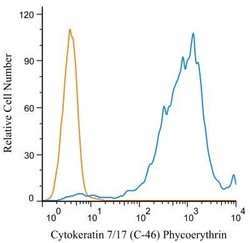
- Flow Cytometry: Cytokeratin 7/17 Antibody (C-46) [NB500-352] - An intracellular stain was performed on HeLa cells with Cytokeratin 7/17 antibody (C-46) NB500-352PE (blue) and a matched isotype control (orange). Cells were fixed with 4% PFA and then permeablized with 0.1% saponin. Cells were incubated in an antibody dilution of 1 ug/mL for 30 minutes at room temperature. Both antibodies were conjugated to Phycoerythrin.

- Submitted by
- Novus Biologicals (provider)
- Main image

- Experimental details
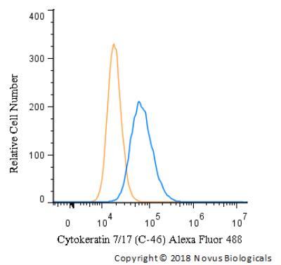
- Flow Cytometry: Cytokeratin 7/17 Antibody (C-46) [NB500-352] - Using the Alexa Fluor 488 direct conjugate An intracellular stain was performed on A549 cells with Cytokeratin 7/17 (C-46) antibody NB500-352AF488 (blue) and a matched isotype control (orange). Cells were fixed with 4% PFA and then permeablized with 0.1% saponin. Cells were incubated in an antibody dilution of 5 ug/mL for 30 minutes at room temperature. Both antibodies were conjugated to Alexa Fluor 488.
- Submitted by
- Novus Biologicals (provider)
- Main image

- Experimental details
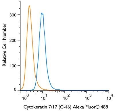
- Flow (Intracellular): Cytokeratin 7/17 Antibody (C-46) [NB500-352] - An intracellular stain was performed on HeLa cells with Cytokeratin 7/17 (C-46) antibody NB500-352AF488 (blue) and a matched isotype control (orange). Cells were fixed with 4% PFA and then permeablized with 0.1% saponin. Cells were incubated in an antibody dilution of 5 ug/mL for 30 minutes at room temperature. Both antibodies were conjugated to Alexa Fluor 488.