Explore
 Validate
 Learn
Learn Western blot
Antibody data
- Antibody Data
- Antigen structure
- References [4]
- Comments [0]
- Validations
- Western blot [1]
- Immunocytochemistry [2]
Submit
Validation data
Reference
Comment
Report error
- Product number
- 14-9005-80 - Provider product page

- Provider
- Invitrogen Antibodies
- Product name
- Cytokeratin 7 Monoclonal Antibody (LP5K), eBioscience™
- Antibody type
- Monoclonal
- Antigen
- Other
- Description
- Description: This LP5K monoclonal antibody reacts with human cytokeratin 7 (K7), a 54-kDa type II (or basic) keratin expressed either alone or paired with cytokeratin 19 in simple epithelia, mesothelium, urothelium, and pseudostratified epithelium. Expression of cytokeratin 7 in the gastric foveolar, intestinal, and stratified squamous epithelia is extremely low or undetectable. Cytokeratins form the intracellular cytoskeletal network that maintains the integrity and stability of cells and tissues. In addition, most carcinomas express cytokeratin 7. The coordinated expression of this keratin with cytokeratin 20 is commonly used as a diagnostic marker for a variety of carcinomas. Applications Reported: This LP5K antibody has been reported for use in immunoblotting (WB) and immunocytochemistry (ICC). Applications Tested: This LP5K antibody has been tested by immunofluorescent staining of paraformaldehyde fixed and permeabilized cells. This can be used at less than or equal to 10 µg/mL. It is recommended that the antibody be titrated for optimal performance in the assay of interest. Purity: Greater than 90%, as determined by SDS-PAGE. Aggregation: Less than 10%, as determined by HPLC. Filtration: 0.2 µm post-manufacturing filtered.
- Reactivity
- Human
- Host
- Mouse
- Isotype
- IgG
- Antibody clone number
- LP5K
- Vial size
- 25 µg
- Concentration
- 0.5 mg/mL
- Storage
- 4° C
Submitted references CCNE1 copy number is a biomarker for response to combination WEE1-ATR inhibition in ovarian and endometrial cancer models.
The human keratins: biology and pathology.
Platelet-derived soluble factors induce human extravillous trophoblast migration and differentiation: platelets are a possible regulator of trophoblast infiltration into maternal spiral arteries.
Cytokeratin 7 and cytokeratin 20 expression in epithelial neoplasms: a survey of 435 cases.
Xu H, George E, Kinose Y, Kim H, Shah JB, Peake JD, Ferman B, Medvedev S, Murtha T, Barger CJ, Devins KM, D'Andrea K, Wubbenhorst B, Schwartz LE, Hwang WT, Mills GB, Nathanson KL, Karpf AR, Drapkin R, Brown EJ, Simpkins F
Cell reports. Medicine 2021 Sep 21;2(9):100394
Cell reports. Medicine 2021 Sep 21;2(9):100394
The human keratins: biology and pathology.
Moll R, Divo M, Langbein L
Histochemistry and cell biology 2008 Jun;129(6):705-33
Histochemistry and cell biology 2008 Jun;129(6):705-33
Platelet-derived soluble factors induce human extravillous trophoblast migration and differentiation: platelets are a possible regulator of trophoblast infiltration into maternal spiral arteries.
Sato Y, Fujiwara H, Zeng BX, Higuchi T, Yoshioka S, Fujii S
Blood 2005 Jul 15;106(2):428-35
Blood 2005 Jul 15;106(2):428-35
Cytokeratin 7 and cytokeratin 20 expression in epithelial neoplasms: a survey of 435 cases.
Chu P, Wu E, Weiss LM
Modern pathology : an official journal of the United States and Canadian Academy of Pathology, Inc 2000 Sep;13(9):962-72
Modern pathology : an official journal of the United States and Canadian Academy of Pathology, Inc 2000 Sep;13(9):962-72
No comments: Submit comment
Supportive validation
- Submitted by
- Invitrogen Antibodies (provider)
- Main image

- Experimental details
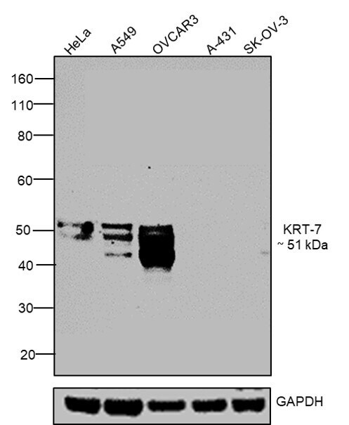
- Western blot was performed using Cytokeratin 7 Monoclonal Antibody (LP5K), eBioscience™ (Product # 14-9005-82) and a 51 kDa band corresponding to KRT-7 was observed in positive cell lines such as HeLa, A549, OVCAR-3 and not in negative cell lines such as A431 and SK-OV-3 as mentioned in literature. Membrane enriched extracts (30 µg lysate) of HeLa (Lane 1), A549 (Lane 2), OVCAR-3 (Lane 3), A-431 (Lane 4) and SK-OV-3 (Lane 5) were electrophoresed using NuPAGE® 4-12 % Bis-Tris gel (Product # NP0321BOX). Resolved proteins were then transferred onto a nitrocellulose membrane (Product # IB23001) by iBlot® 2 Dry Blotting System (Product # IB21001). The blot was probed with the primary antibody (1:1000 dilution) and detected by chemiluminescence with Goat anti-Mouse IgG (H+L) Superclonal™ Recombinant Secondary Antibody, HRP (Product # A28177, 1:4000 dilution) using the iBright FL 1000 (Product # A32752). Chemiluminescent detection was performed using Novex® ECL Chemiluminescent Substrate Reagent Kit (Product # WP20005).
Supportive validation
- Submitted by
- Invitrogen Antibodies (provider)
- Main image

- Experimental details
- Immunocytochemistry of fixed and permeabilized HeLa cells using 10 µg/mL of Mouse IgG2b Isotype Control (Product # 14-4732-82) (left) or Anti-Human Cytokeratin 7 Purified (right) followed by Anti-Mouse TRITC. Nuclei are counterstained with DAPI.

- Submitted by
- Invitrogen Antibodies (provider)
- Main image
- Experimental details
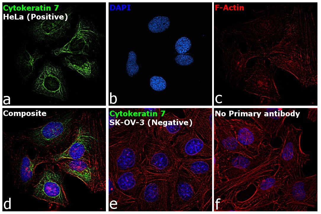
- Immunofluorescence analysis of Cytokeratin 7 was performed using HeLa and SK-OV-3 cells. The cells were fixed with 4% paraformaldehyde for 10 minutes, permeabilized with 0.1% Triton™ X-100 for 15 minutes, and blocked with 2% BSA for 1 hour at room temperature. The cells were labeled with Cytokeratin 7 Monoclonal Antibody (LP5K), eBioscience™ (Product # 14-9005-82) at 5 microgram/mL in 0.1% BSA, incubated at 4 degree celsius overnight and then labeled with Goat anti-Mouse IgG (H+L) Superclonal™ Recombinant Secondary Antibody, Alexa Fluor® 488 (Product # A28175) at a dilution of 1:2000 for 45 minutes at room temperature (Panel a: green). Nuclei (Panel b: blue) were stained with ProLong™ Diamond Antifade Mountant with DAPI (Product # P36962). F-actin (Panel c: red) was stained with Rhodamine Phalloidin (Product # R415, 1:300). Panel d represents the merged image showing expression of Cytokeratin 7 in HeLa. Panel e represents SK-OV-3 cells, showing lesser expression of Cytokeratin 7. Panel f represents control HeLa cells with no primary antibody to assess background. The images were captured at 60X magnification.