 Explore
 Validate
 Learn
Learn Western blot
 ELISA
ELISAAntibody data
- Antibody Data
- Antigen structure
- References [1]
- Comments [0]
- Validations
- Western blot [4]
- Immunocytochemistry [1]
- Flow cytometry [1]
Submit
Validation data
Reference
Comment
Report error
- Product number
- MA5-15604 - Provider product page

- Provider
- Invitrogen Antibodies
- Product name
- Cytokeratin 7 Monoclonal Antibody (5D12)
- Antibody type
- Monoclonal
- Antigen
- Purifed from natural sources
- Description
- MA5-15604 targets CK7 in indirect ELISA, FACS and WB applications and shows reactivity with Human samples. The MA5-15604 immunogen is purified recombinant fragment of human CK7 expressed in E. Coli. MA5-15604 detects CK7 which has a predicted molecular weight of approximately 51kDa.
- Reactivity
- Human
- Host
- Mouse
- Isotype
- IgG
- Antibody clone number
- 5D12
- Vial size
- 100 µL
- Concentration
- Conc. Not Determined
- Storage
- Store at 4°C short term. For long term storage, store at -20°C, avoiding freeze/thaw cycles.
Submitted references Detection and analysis of multiple biomarkers in ovarian cancer: clinical significance in diagnosis, treatment, and prognosis evaluation.
Ji R, Li Y, He C, Zhu X, He A, Lu Y
Gland surgery 2020 Dec;9(6):2175-2186
Gland surgery 2020 Dec;9(6):2175-2186
No comments: Submit comment
Supportive validation
- Submitted by
- Invitrogen Antibodies (provider)
- Main image

- Experimental details
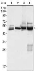
- Western blot analysis of CK7 using CK7 monoclonal antibody (Product # MA5-15604) in HeLa (1), MCF-7 (2), A431 (3) and A549 (4) cell lysate.

- Submitted by
- Invitrogen Antibodies (provider)
- Main image
- Experimental details
- Western blot analysis of CK7 using CK7 monoclonal antibody (Product # MA5-15604) in HeLa (1), MCF-7 (2), A431 (3) and A549 (4) cell lysate.

- Submitted by
- Invitrogen Antibodies (provider)
- Main image

- Experimental details
- Western blot analysis of CK7 using CK7 monoclonal antibody (Product # MA5-15604) in HeLa (1), MCF-7 (2), A431 (3) and A549 (4) cell lysate.

- Submitted by
- Invitrogen Antibodies (provider)
- Main image
- Experimental details
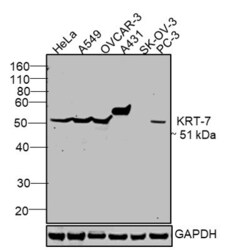
- Western blot was performed using Anti-Cytokeratin 7 Monoclonal Antibody (5D12) (Product # MA5-15604) and a 51 kDa band corresponding to KRT-7 was observed in positive cell lines like HeLa, A549, OVCAR-3, A-431, PC-3 and not in negative cell lines live SK-OV-3 as mentioned in literature. Membrane enriched extracts (30 µg lysate) of HeLa (Lane 1), A549 (Lane 2), OVCAR-3 (Lane 3), A-431 (Lane 4), SK-OV-3 (Lane 5) and PC-3 (Lane 6) were electrophoresed using NuPAGE® 4-12 % Bis-Tris gel (Product # NP0321BOX). Resolved proteins were then transferred onto a nitrocellulose membrane (Product # IB23001) by iBlot® 2 Dry Blotting System (Product # IB21001). The blot was probed with the primary antibody (1:1000 dilution) and detected by chemiluminescence with Goat anti-Mouse IgG (H+L), Superclonal™ Recombinant Secondary Antibody, HRP conjugate (Product # A28177, 1:4000 dilution) using the iBright FL 1000 (Product # A32752). Chemiluminescent detection was performed using Novex® ECL Chemiluminescent Substrate Reagent Kit (Product # WP20005).

Supportive validation
- Submitted by
- Invitrogen Antibodies (provider)
- Main image

- Experimental details
- Immunofluorescence analysis of Cytokeratin 7 was performed using 70% confluent log phase HeLa cells. The cells were fixed and permeabilized with ice cold 100% Acetone for 5 minutes, and blocked with 2% BSA for 1 hour at room temperature. The cells were labeled with Cytokeratin 7 Monoclonal Antibody (5D12) (Product # MA5-15604) at 1:200 dilution in 0.1% BSA, incubated at 4 degree Celsius overnight and then labeled with Goat anti-Mouse IgG (H+L) Superclonal™ Recombinant Secondary Antibody, Alexa Fluor® 488 conjugate (Product # A28175) at a dilution of 1:2000 for 45 minutes at room temperature (Panel a: green). Nuclei (Panel b: blue) were stained with ProLong™ Diamond Antifade Mountant with DAPI (Product # P36962). F-actin (Panel c: red) was stained with Rhodamine Phalloidin (Product # R415, 1:300). Panel d represents the merged image showing cytoplasmic localization. Panel e represents control cells with no primary antibody to assess background. The images were captured at 60X magnification.

Supportive validation
- Submitted by
- Invitrogen Antibodies (provider)
- Main image

- Experimental details
- Flow cytometric analysis of HeLa cells using CK7 monoclonal antibody (Product # MA5-15604) (green) and negative control (purple).
