 Explore
 Validate
 Learn
Learn Western blot
 Immunocytochemistry
Antibody data
- Antibody Data
- Antigen structure
- References [2]
- Comments [0]
- Validations
- Immunocytochemistry [4]
- Immunohistochemistry [1]
- Flow cytometry [1]
Submit
Validation data
Reference
Comment
Report error
- Product number
- 701989 - Provider product page

- Provider
- Invitrogen Antibodies
- Product name
- Glutamine Synthetase Recombinant Rabbit Monoclonal Antibody (7H9L16)
- Antibody type
- Monoclonal
- Antigen
- Other
- Description
- This antibody is predicted to react with Monkey, Cat and Pig. Recombinant rabbit monoclonal antibodies are produced using in vitro expression systems. The expression systems are developed by cloning in the specific antibody DNA sequences from immunoreactive rabbits. Then, individual clones are screened to select the best candidates for production. The advantages of using recombinant rabbit monoclonal antibodies include: better specificity and sensitivity, lot-to-lot consistency, animal origin-free formulations, and broader immunoreactivity to diverse targets due to larger rabbit immune repertoire.
- Reactivity
- Human, Mouse, Rat
- Host
- Rabbit
- Isotype
- IgG
- Antibody clone number
- 7H9L16
- Vial size
- 100 μg
- Concentration
- 0.5 mg/mL
- Storage
- Store at 4°C short term. For long term storage, store at -20°C, avoiding freeze/thaw cycles.
Submitted references Extracellular Vesicles From 3xTg-AD Mouse and Alzheimer's Disease Patient Astrocytes Impair Neuroglial and Vascular Components.
Cellular and molecular outcomes of glutamine supplementation in the brain of succinic semialdehyde dehydrogenase-deficient mice.
González-Molina LA, Villar-Vesga J, Henao-Restrepo J, Villegas A, Lopera F, Cardona-Gómez GP, Posada-Duque R
Frontiers in aging neuroscience 2021;13:593927
Frontiers in aging neuroscience 2021;13:593927
Cellular and molecular outcomes of glutamine supplementation in the brain of succinic semialdehyde dehydrogenase-deficient mice.
Brown MN, Gibson KM, Schmidt MA, Walters DC, Arning E, Bottiglieri T, Roullet JB
JIMD reports 2020 Nov;56(1):58-69
JIMD reports 2020 Nov;56(1):58-69
No comments: Submit comment
Supportive validation
- Submitted by
- Invitrogen Antibodies (provider)
- Main image
- Experimental details
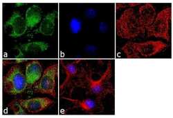
- Immunofluorescence was performed on methanol fixed HepG2 cells for detection of endogenous Glutaminase synthetase using Glutaminase synthetase Recombinant Rabbit Monoclonal Antibody (Product # 701989, 2 µg/mL) and labeled with Goat anti-Rabbit IgG (H+L) Superclonal™ Secondary Antibody, Alexa Fluor® 488 conjugate (Product # A27034, 1:2000). Cytoskeleton was stained with alpha-Tubulin Monoclonal Antibody (Product # 32-2500, 1 µg/mL) followed by Goat anti-Mouse IgG Secondary Antibody, Alexa Fluor® 594 conjugate (Product # A-11032, 1:400). Panel a) shows representative cells that were stained for detection and localization of Glutaminase synthetase protein (green), Panel b) is stained for nuclei (blue) using SlowFade® Gold Antifade Mountant with DAPI (Product # S36938). Panel c) represents cytoskeletal alpha-tubulin staining (red). Panel d) is a composite image of Panels a, b and c clearly demonstrating cytoplasmic localization of Glutaminase synthetase. Panel e) represents control cells with no primary antibody to assess background. The images were captured at 60X magnification.

- Submitted by
- Invitrogen Antibodies (provider)
- Main image
- Experimental details
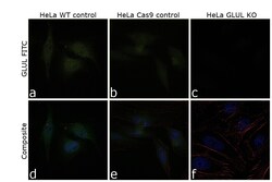
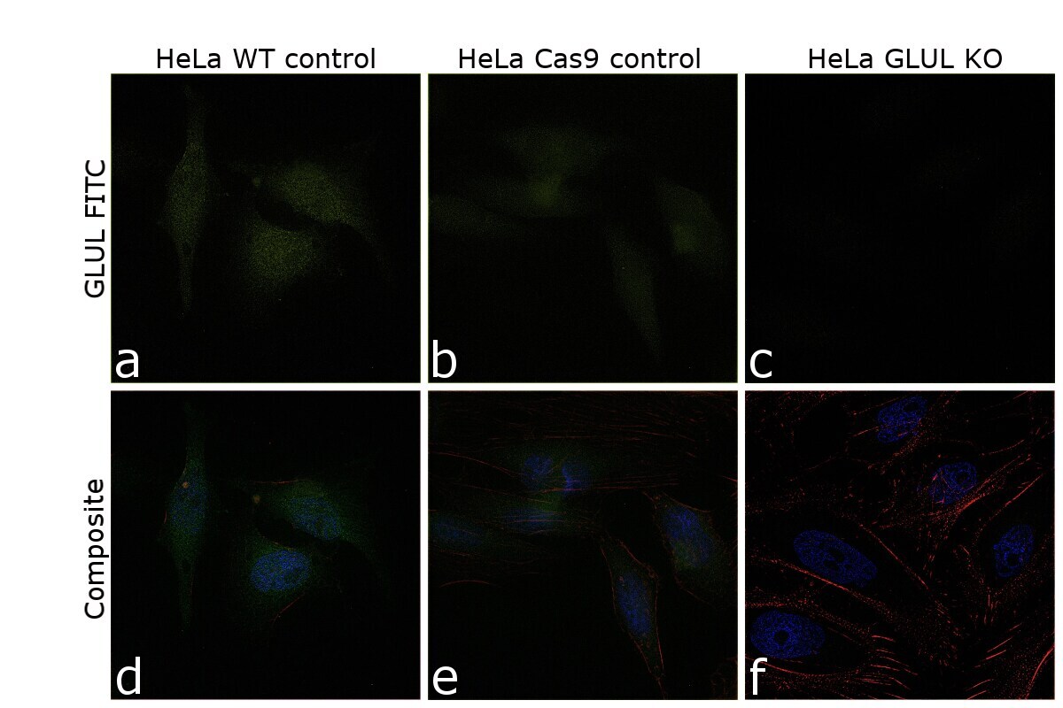
- Knockout of Glutamine Synthetase was achieved by CRISPR-Cas9 genome editing using LentiArray™ Lentiviral sgRNA (Product # A32042, AssayID CRISPR531952_LV) and LentiArray Cas9 Lentivirus (Product # A32064). Immunofluorescence analysis was performed on wild type HeLa cells (panel a,d), HeLa Cas9 cells (panels b,e) and HeLa Glutamine Synthetase KO cells (panel c,f). Cells were fixed, permeabilized, and labelled with Anti-Glutamine Synthetase Recombinant Rabbit Monoclonal Antibody (7H9L16) (Product # 7019898203, 2µg/mL), followed by Goat anti-Rabbit IgG (H+L) Highly Cross-Adsorbed Secondary Antibody, Alexa Fluor Plus 488 (Product # A32731TR, 1:2000). Nuclei (blue) were stained using ProLong™ Diamond Antifade Mountant with DAPI (Product # P36962), and Rhodamine Phalloidin (Product # R415, 1:300) was used for cytoskeletal F-actin (red) staining. Loss of signal (panel c,f) upon CRISPR mediated knockout (KO) confirms that antibody is specific to Glutamine Synthetase (green). The images were captured at 60X magnification.
- Submitted by
- Invitrogen Antibodies (provider)
- Main image

- Experimental details
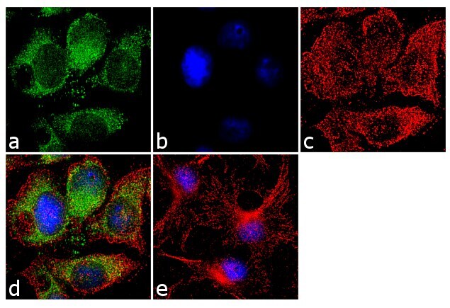
- Immunofluorescence was performed on methanol fixed HepG2 cells for detection of endogenous Glutaminase synthetase using Glutaminase synthetase Recombinant Rabbit Monoclonal Antibody (Product # 701989, 2 µg/mL) and labeled with Goat anti-Rabbit IgG (Heavy Chain) Superclonal™ Secondary Antibody, Alexa Fluor® 488 conjugate (Product # A27034, 1:2000). Cytoskeleton was stained with alpha-Tubulin Monoclonal Antibody (Product # 32-2500, 1 µg/mL) followed by Goat anti-Mouse IgG Secondary Antibody, Alexa Fluor® 594 conjugate (Product # A-11032, 1:400). Panel a) shows representative cells that were stained for detection and localization of Glutaminase synthetase protein (green), Panel b) is stained for nuclei (blue) using SlowFade® Gold Antifade Mountant with DAPI (Product # S36938). Panel c) represents cytoskeletal alpha-tubulin staining (red). Panel d) is a composite image of Panels a, b and c clearly demonstrating cytoplasmic localization of Glutaminase synthetase. Panel e) represents control cells with no primary antibody to assess background. The images were captured at 60X magnification.
- Submitted by
- Invitrogen Antibodies (provider)
- Main image

- Experimental details
- Knockout of Glutamine Synthetase was achieved by CRISPR-Cas9 genome editing using LentiArray™ Lentiviral sgRNA (Product # A32042, AssayID CRISPR531952_LV) and LentiArray Cas9 Lentivirus (Product # A32064). Immunofluorescence analysis was performed on wild type HeLa cells (panel a,d), HeLa Cas9 cells (panels b,e) and HeLa Glutamine Synthetase KO cells (panel c,f). Cells were fixed, permeabilized, and labelled with Anti-Glutamine Synthetase Recombinant Rabbit Monoclonal Antibody (7H9L16) (Product # 7019898203, 2µg/mL), followed by Goat anti-Rabbit IgG (H+L) Highly Cross-Adsorbed Secondary Antibody, Alexa Fluor Plus 488 (Product # A32731TR, 1:2000). Nuclei (blue) were stained using ProLong™ Diamond Antifade Mountant with DAPI (Product # P36962), and Rhodamine Phalloidin (Product # R415, 1:300) was used for cytoskeletal F-actin (red) staining. Loss of signal (panel c,f) upon CRISPR mediated knockout (KO) confirms that antibody is specific to Glutamine Synthetase (green). The images were captured at 60X magnification.

Supportive validation
- Submitted by
- Invitrogen Antibodies (provider)
- Main image
- Experimental details
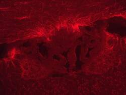
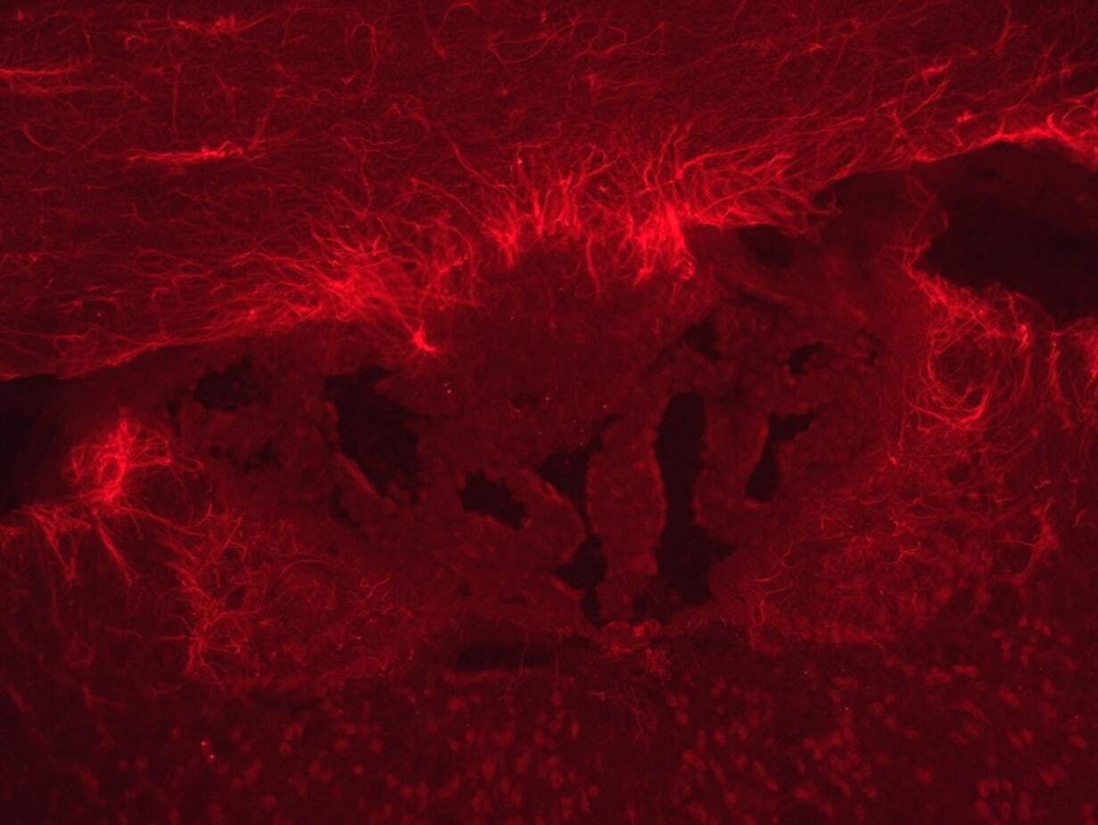
- Immunofluorescence analysis of glutamine synthetase (red) in mouse brain cortex tissue. The tissues were fixed with 4% PFA for 1 week, there is no permeabilization process because of frozen section, and blocked with 3% donkey serum for 1hr in RT. Tissues were stained with a recombinant monoclonal glutamine synthetase antibody (Product # 701989) at a dilution of 1:1000 in PBS for overnight in 4°C, and then incubated with secondary donkey anti-rabbit IgG (H+L)- Alexa Fluor 594 antibody (Product # A-21207) at a dilution of 1:500 for 1hour. Image was taken at 20X magnification. Data courtesy of Cherl Namkoong at Seoul National University, Korea.
Supportive validation
- Submitted by
- Invitrogen Antibodies (provider)
- Main image
- Experimental details
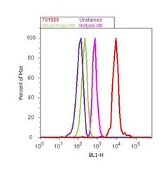
- Flow Cytometry analysis of endogenous Glutaminase synthetase was performed on Hep G2 cells labeled with ABfinity™ Anti-Glutaminase synthetase Recombinant Rabbit Monoclonal Antibody (Product# 701989, 5 ug/ 1M cells) or with rabbit isotype control at 0.5 ug/ml and detected with Goat anti-Rabbit IgG (H+L) Superclonal™ Secondary Antibody, (Alexa Fluor® 488 conjugate, Product# A27034, 0.4 ug/ml, 1:2500) as represented by the red and pink histograms respectively. The purple histogram represents unstained control cells and the green histogram represents no-primary-antibody control. A representative of 10,000 cells were acquired and analyzed for each sample using an Attune® Acoustic Focusing Cytometer (4468770).
