Explore
 Validate
 Learn
Learn Western blot
Antibody data
- Antibody Data
- Antigen structure
- References [0]
- Comments [0]
- Validations
- Western blot [2]
- Immunocytochemistry [1]
Submit
Validation data
Reference
Comment
Report error
- Product number
- 710963 - Provider product page

- Provider
- Invitrogen Antibodies
- Product name
- Glutamine Synthetase Recombinant Polyclonal Antibody (7HCLC)
- Antibody type
- Polyclonal
- Antigen
- Other
- Reactivity
- Human
- Host
- Rabbit
- Isotype
- IgG
- Antibody clone number
- 7HCLC
- Vial size
- 100 µg
- Concentration
- 0.5 mg/mL
- Storage
- Store at 4°C short term. For long term storage, store at -20°C, avoiding freeze/thaw cycles.
No comments: Submit comment
Supportive validation
- Submitted by
- Invitrogen Antibodies (provider)
- Main image

- Experimental details
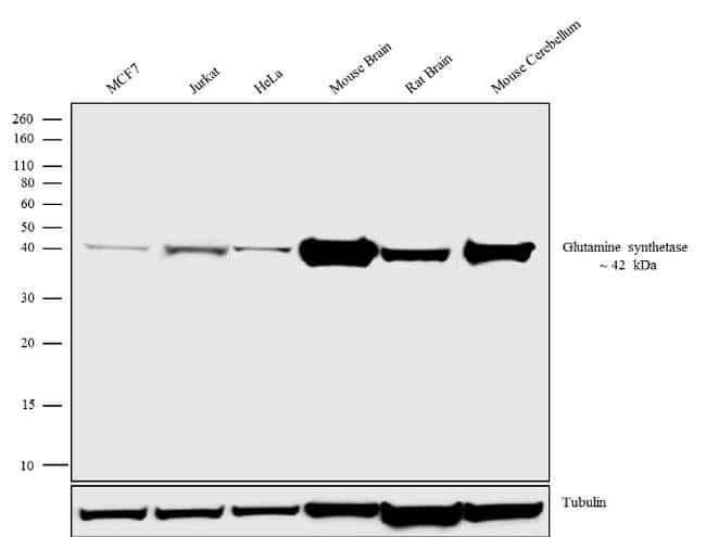
- Western blot analysis was performed on whole cell extracts (30 µg lysate) of MCF7 (Lane 1), Jurkat (Lane 2), HeLa (Lane 3) and tissue extracts of Mouse Brain (Lane 4), Rat Brain (Lane 5) and Mouse Cerebellum (Lane 6). The blots were probed with Anti-Glutamine synthetase Recombinant Rabbit Monoclonal Antibody (Product # 710963, 1-2 µg/mL) and detected by chemiluminescence using Goat anti-Rabbit IgG (H+L) Superclonal™ Secondary Antibody, HRP conjugate (Product # A27036, 0.4 µg/mL, 1:2500 dilution). A 42 kDa band corresponding to Glutamine synthetase was observed. Known quantity of protein samples were electrophoresed using Novex® NuPAGE® 10% Bis-Tris gel (Product # NP0301BOX), XCell SureLock™ Electrophoresis System (Product # EI0002) and Novex® Sharp Pre-Stained Protein Standard (Product # LC5800). Resolved proteins were then transferred onto a nitrocellulose membrane with iBlot® Dry Blotting System (Product # IB21001). The membrane was probed with the relevant primary and secondary Antibody following blocking with 5% skimmed milk. Chemiluminescent detection was performed using Pierce™ ECL Western blotting Substrate (Product # 32106).
- Submitted by
- Invitrogen Antibodies (provider)
- Main image

- Experimental details
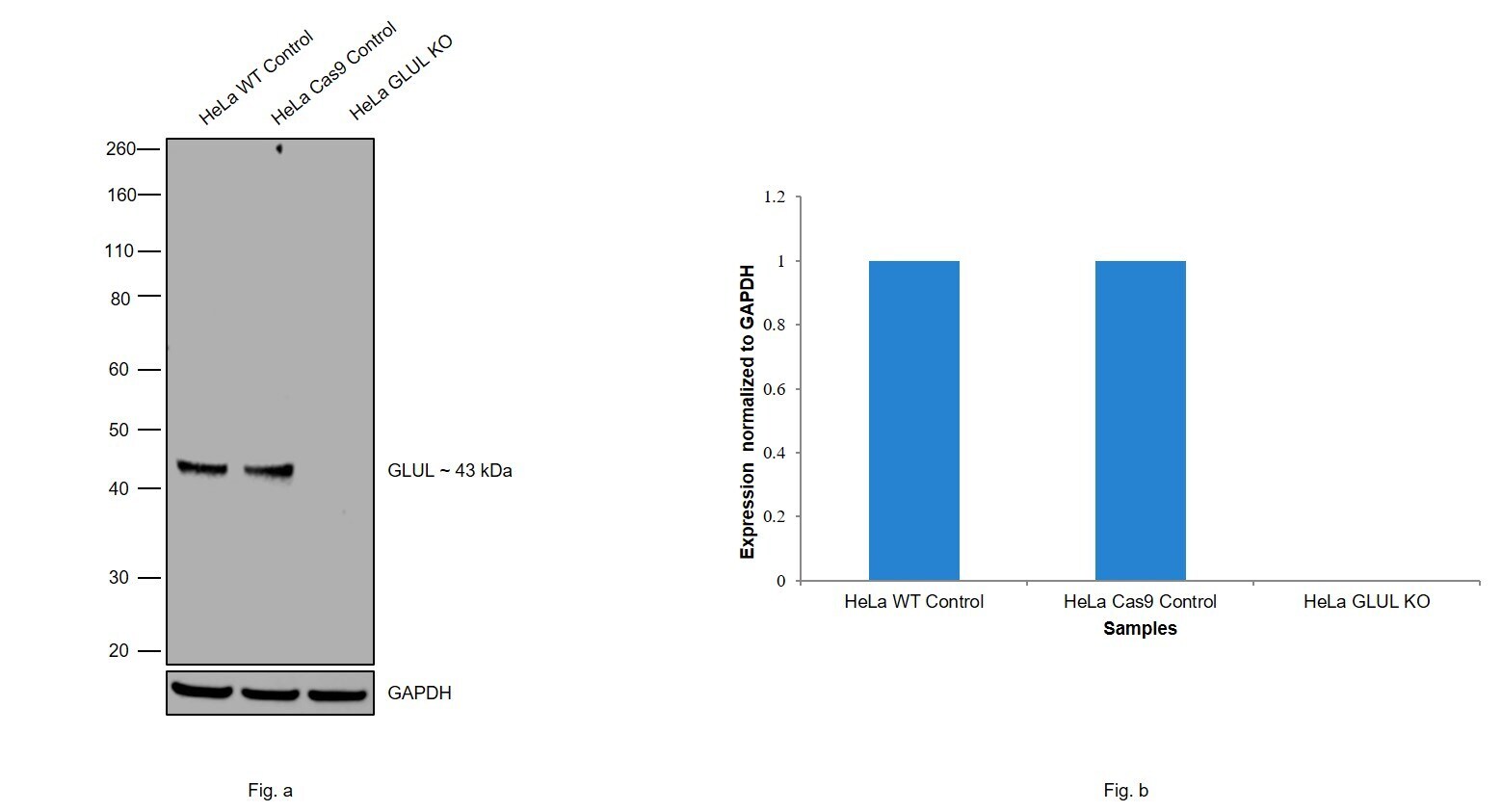
- Knockout of Glutamine Synthetase was achieved by CRISPR-Cas9 genome editing using LentiArray™ Lentiviral sgRNA (Product # A32042, AssayID CRISPR531952_LV) and LentiArray Cas9 Lentivirus (Product # A32064). Western blot analysis of Glutamine Synthetase was performed by loading 20 µg of HeLa wild type (Lane 1), HeLa CAS9 (Lane 2), HeLa Glutamine Synthetase KO (Lane 3) whole cell extracts. The blot was probed with Anti-Glutamine Synthetase Recombinant Polyclonal Antibody (7HCLC)(Product # 710963) using 2 µg/mL dilution and Goat anti-Rabbit IgG (H+L), Superclonal™ Recombinant Secondary Antibody, HRP (Product # A27036). Loss of signal upon CRISPR mediated knockout (KO) confirms that the antibody is specific to Glutamine Synthetase.
Supportive validation
- Submitted by
- Invitrogen Antibodies (provider)
- Main image
- Experimental details
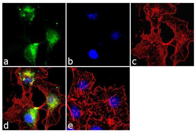
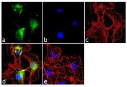
- Immunofluorescence was performed on fixed and permeabilized HepG2 cells for detection of Glutamine synthetase using Glutamine synthetase Recombinant Rabbit Polyclonal Antibody (Product # 710963, 2 µg/mL) and labeled with Goat anti-Rabbit IgG (H+L) Superclonal™ Secondary Antibody, Alexa Fluor® 488 conjugate (Product # A27034, 1:2000). Panel a) shows representative cells that were stained for detection and localization of Glutamine synthetase protein (green), Panel b) is stained for nuclei (blue) using SlowFade® Gold Antifade Mountant with DAPI (Product # S36938). Panel c) represents cytoskeletal F-actin staining using Alexa Fluor® 555 Rhodamine Phalloidin (Product # R415, 1:300). Panel d) is a composite image of Panels a, b and c clearly demonstrating cytoplasmic localization of Glutamine synthetase. Panel e) represents control cells with no primary antibody to assess background.