Explore
 Validate
 Learn
Learn Western blot
 Immunohistochemistry
Antibody data
- Antibody Data
- Antigen structure
- References [5]
- Comments [0]
- Validations
- Immunohistochemistry [1]
- Other assay [1]
Submit
Validation data
Reference
Comment
Report error
- Product number
- PA1-46165 - Provider product page

- Provider
- Invitrogen Antibodies
- Product name
- Glutamine Synthetase Polyclonal Antibody
- Antibody type
- Polyclonal
- Antigen
- Recombinant full-length protein
- Description
- Suggested positive control: antigen standard for GLUL (transient overexpression lysate), retinal muller cells.
- Reactivity
- Human, Mouse, Rat, Bovine
- Host
- Rabbit
- Isotype
- IgG
- Vial size
- 100 μL
- Concentration
- Conc. Not Determined
- Storage
- -20°C or -80°C if preferred
Submitted references VEGF Mediates Retinal Müller Cell Viability and Neuroprotection through BDNF in Diabetes.
Tissue-Specific Regulation of the Wnt/β-Catenin Pathway by PAGE4 Inhibition of Tankyrase.
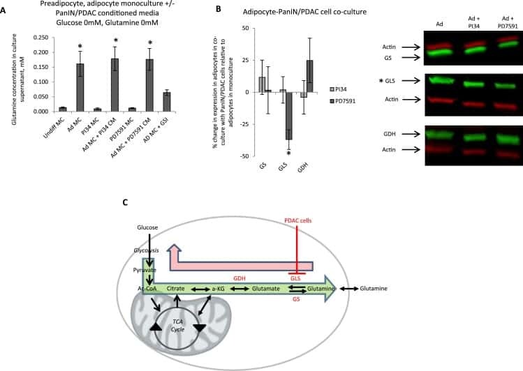
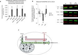
Adipocytes promote pancreatic cancer cell proliferation via glutamine transfer.
Neurovascular crosstalk between interneurons and capillaries is required for vision.
Defective retinal vascular endothelial cell development as a consequence of impaired integrin αVβ8-mediated activation of transforming growth factor-β.
Le YZ, Xu B, Chucair-Elliott AJ, Zhang H, Zhu M
Biomolecules 2021 May 10;11(5)
Biomolecules 2021 May 10;11(5)
Tissue-Specific Regulation of the Wnt/β-Catenin Pathway by PAGE4 Inhibition of Tankyrase.
Koirala S, Klein J, Zheng Y, Glenn NO, Eisemann T, Fon Tacer K, Miller DJ, Kulak O, Lu M, Finkelstein DB, Neale G, Tillman H, Vogel P, Strand DW, Lum L, Brautigam CA, Pascal JM, Clements WK, Potts PR
Cell reports 2020 Jul 21;32(3):107922
Cell reports 2020 Jul 21;32(3):107922
Adipocytes promote pancreatic cancer cell proliferation via glutamine transfer.
Meyer KA, Neeley CK, Baker NA, Washabaugh AR, Flesher CG, Nelson BS, Frankel TL, Lumeng CN, Lyssiotis CA, Wynn ML, Rhim AD, O'Rourke RW
Biochemistry and biophysics reports 2016 Sep;7:144-149
Biochemistry and biophysics reports 2016 Sep;7:144-149
Neurovascular crosstalk between interneurons and capillaries is required for vision.
Usui Y, Westenskow PD, Kurihara T, Aguilar E, Sakimoto S, Paris LP, Wittgrove C, Feitelberg D, Friedlander MS, Moreno SK, Dorrell MI, Friedlander M
The Journal of clinical investigation 2015 Jun;125(6):2335-46
The Journal of clinical investigation 2015 Jun;125(6):2335-46
Defective retinal vascular endothelial cell development as a consequence of impaired integrin αVβ8-mediated activation of transforming growth factor-β.
Arnold TD, Ferrero GM, Qiu H, Phan IT, Akhurst RJ, Huang EJ, Reichardt LF
The Journal of neuroscience : the official journal of the Society for Neuroscience 2012 Jan 25;32(4):1197-206
The Journal of neuroscience : the official journal of the Society for Neuroscience 2012 Jan 25;32(4):1197-206
No comments: Submit comment
Supportive validation
- Submitted by
- Invitrogen Antibodies (provider)
- Main image
- Experimental details
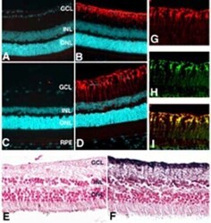
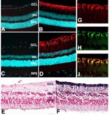
- Immunohistochemical analysis of Glutamine Synthetase in Paraffin sections of mouse (A, B), rat (C, D, G-I), or human (E, F) retina fixed in 4% paraformaldehyde. Samples were incubated in Glutamine Synthetase polyclonal antibody (Product # PA1-46165). Anti-glutamine synthase (red fluorescence staining in B, D, G, I, and brown immuno-peroxidase reaction. Nuclei in IF experiments (A-D) were stained with DAPI (cyan), and with nuclear fast red in E and F. At low magnification, anti-glutamine synthase reacted with a single population of cells extending from the ganglion cell layer through the inner nuclear layer. No signal was detected in controls either pre-incubated with 100 µg/mL of the immunizing peptide (A) or with pre-immune serum (C, E). This finding was confirmed by co-localization (indicated by yellow in I) of glutamine synthase (red in G) with antoher marker of glutamine synthase (green in H).
Supportive validation
- Submitted by
- Invitrogen Antibodies (provider)
- Main image
- Experimental details
- NULL