 Explore
 Validate
 Learn
Learn Western blot
 Immunocytochemistry
Antibody data
- Antibody Data
- Antigen structure
- References [5]
- Comments [0]
- Validations
- Immunocytochemistry [2]
- Immunoprecipitation [1]
- Immunohistochemistry [3]
- Other assay [1]
Submit
Validation data
Reference
Comment
Report error
- Product number
- PA5-28940 - Provider product page

- Provider
- Invitrogen Antibodies
- Product name
- Glutamine Synthetase Polyclonal Antibody
- Antibody type
- Polyclonal
- Antigen
- Recombinant full-length protein
- Description
- Recommended positive controls: 293T, U87-MG, SK-N-SH, IMR32, SK-N-AS, mouse brain, mouse eye, rat brain. Predicted reactivity: Mouse (94%), Rat (92%), Xenopus laevis (85%), Dog (97%), Pig (96%), Chicken (88%), Rhesus Monkey (100%), Chimpanzee (99%), Bovine (96%). Store product as a concentrated solution. Centrifuge briefly prior to opening the vial.
- Reactivity
- Human, Mouse, Rat
- Host
- Rabbit
- Isotype
- IgG
- Vial size
- 100 μL
- Concentration
- 0.79 mg/mL
- Storage
- Store at 4°C short term. For long term storage, store at -20°C, avoiding freeze/thaw cycles.
Submitted references Phenotypical changes of satellite glial cells in a murine model of G(M1) -gangliosidosis.
Immunohistochemical Characterization of the Nervous System of Culex pipiens (Diptera, Culicidae).
Analysis of mir-9 Expression Pattern in Rat Retina during Postnatal Development.
Phenotypical peculiarities and species-specific differences of canine and murine satellite glial cells of spinal ganglia.
Glutamine synthetase in avian muscle contributes to a positive myogenic response to ammonia compared with mammalian muscle.
Huang B, Zdora I, de Buhr N, Eikelberg D, Baumgärtner W, Leitzen E
Journal of cellular and molecular medicine 2022 Jan;26(2):527-539
Journal of cellular and molecular medicine 2022 Jan;26(2):527-539
Immunohistochemical Characterization of the Nervous System of Culex pipiens (Diptera, Culicidae).
Gregor KM, Becker SC, Hellhammer F, Baumgärtner W, Puff C
Biology 2022 Jan 1;11(1)
Biology 2022 Jan 1;11(1)
Analysis of mir-9 Expression Pattern in Rat Retina during Postnatal Development.
Pöstyéni E, Kovács-Valasek A, Urbán P, Czuni L, Sétáló G Jr, Fekete C, Gabriel R
International journal of molecular sciences 2021 Mar 4;22(5)
International journal of molecular sciences 2021 Mar 4;22(5)
Phenotypical peculiarities and species-specific differences of canine and murine satellite glial cells of spinal ganglia.
Huang B, Zdora I, de Buhr N, Lehmbecker A, Baumgärtner W, Leitzen E
Journal of cellular and molecular medicine 2021 Jul;25(14):6909-6924
Journal of cellular and molecular medicine 2021 Jul;25(14):6909-6924
Glutamine synthetase in avian muscle contributes to a positive myogenic response to ammonia compared with mammalian muscle.
Stern RA, Mozdziak PE
American journal of physiology. Regulatory, integrative and comparative physiology 2019 Jul 1;317(1):R214-R221
American journal of physiology. Regulatory, integrative and comparative physiology 2019 Jul 1;317(1):R214-R221
No comments: Submit comment
Supportive validation
- Submitted by
- Invitrogen Antibodies (provider)
- Main image

- Experimental details
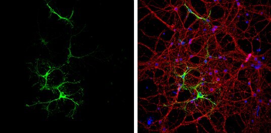
- Immunocytochemistry-Immunofluorescence analysis of Glutamine Synthetase was performed in DIV9 rat E18 primary cortical neurons fixed in 4% paraformaldehyde at RT for 15 min. Green: Glutamine Synthetase Polyclonal Antibody (Product # PA5 28940) diluted at 1:500. Red: beta Tubulin 3/ Tuj1, a neuron cell marker. Blue: Fluoroshield with DAPI.
- Submitted by
- Invitrogen Antibodies (provider)
- Main image

- Experimental details
- Immunocytochemistry-Immunofluorescence analysis of Glutamine Synthetase was performed in DIV9 rat E18 primary cortical neurons fixed in 4% paraformaldehyde at RT for 15 min. Green: Glutamine Synthetase Polyclonal Antibody (Product # PA5 28940) diluted at 1:500. Red: beta Tubulin 3/ Tuj1, a neuron cell marker. Blue: Fluoroshield with DAPI.

Supportive validation
- Submitted by
- Invitrogen Antibodies (provider)
- Main image
- Experimental details
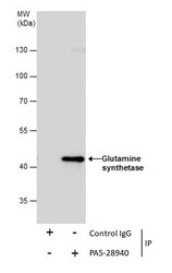
- Immunoprecipitation of Glutamine synthetase was performed in IMR32 whole cell extracts using 5 µg of Glutamine Synthetase Polyclonal Antibody (Product # PA5-28940). Samples were transferred to a membrane and probed with Glutamine Synthetase Polyclonal Antibody as a primary antibody and an HRP-conjugated anti-Rabbit IgG was used as a secondary antibody.
Supportive validation
- Submitted by
- Invitrogen Antibodies (provider)
- Main image
- Experimental details
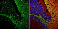
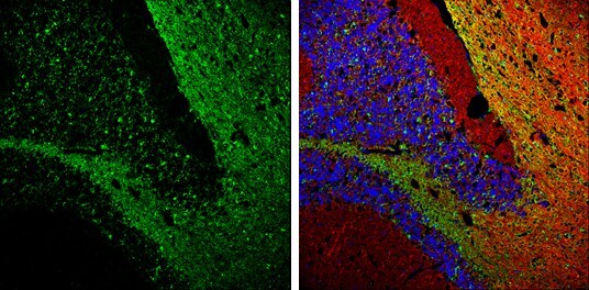
- Immunohistochemistry (Frozen) analysis of Glutamine-Synthetase was performed in frozen-sectioned adult mouse cerebellum tissue using Glutamine Synthetase Polyclonal Antibody (Product # PA5-28940) at a dilution of 1:250 (Green). Red: beta Tubulin 3/ TUJ1, stained by beta Tubulin 3/ TUJ1 antibody diluted at 1:500. Blue: Fluoroshield with DAPI.
- Submitted by
- Invitrogen Antibodies (provider)
- Main image
- Experimental details
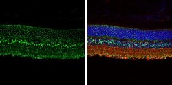
- Immunohistochemistry (Paraffin) analysis of Glutamine Synthetase was performed in paraffin-embedded adult mouse retina tissue using Green: CRX Polyclonal Antibody (Product # PA5-32182) at a dilution of 1:250. Red: beta Tubulin 3/ TUJ1, stained by beta Tubulin 3/ TUJ1 antibody diluted at 1:250. Blue: Fluoroshield with DAPI.

- Submitted by
- Invitrogen Antibodies (provider)
- Main image
- Experimental details
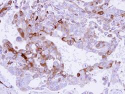
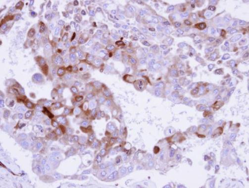
- Immunohistochemical analysis of paraffin-embedded H441 xenograft , using Glutamine Synthetase (Product # PA5-28940) antibody at 1:500 dilution. Antigen Retrieval: EDTA based buffer, pH 8.0, 15 min.
Supportive validation
- Submitted by
- Invitrogen Antibodies (provider)
- Main image

- Experimental details
- Immunoprecipitation of Glutamine synthetase was performed in IMR32 whole cell extracts using 5 µg of Glutamine Synthetase Polyclonal Antibody (Product # PA5-28940). Samples were transferred to a membrane and probed with Glutamine Synthetase Polyclonal Antibody as a primary antibody and an HRP-conjugated anti-Rabbit IgG was used as a secondary antibody.
