Explore
 Validate
 Learn
Learn Western blot
 Immunocytochemistry
Antibody data
- Antibody Data
- Antigen structure
- References [6]
- Comments [0]
- Validations
- Immunocytochemistry [2]
- Immunohistochemistry [1]
Submit
Validation data
Reference
Comment
Report error
- Product number
- MA3-16726 - Provider product page

- Provider
- Invitrogen Antibodies
- Product name
- alpha Internexin Monoclonal Antibody (2E3)
- Antibody type
- Monoclonal
- Antigen
- Purifed from natural sources
- Description
- The epitope recognized by the 2E3 clone is in the C-terminal non-helical extension of the protein and is unusually resistant to aldehyde fixation, so this antibody is ideal for studies of formalin-fixed paraffin-embedded histological sections.
- Reactivity
- Human, Mouse, Rat, Bovine, Porcine
- Host
- Mouse
- Isotype
- IgG
- Antibody clone number
- 2E3
- Vial size
- 100 μL
- Concentration
- 1 mg/mL
- Storage
- Store at 4°C short term. For long term storage, store at -20°C, avoiding freeze/thaw cycles.
Submitted references TBK1 Mutation Spectrum in an Extended European Patient Cohort with Frontotemporal Dementia and Amyotrophic Lateral Sclerosis.
Neuropathological criteria of anti-IgLON5-related tauopathy.
An autopsy case of neuronal intermediate filament inclusion disease with regard to immunophenotypic and topographical analysis of the neuronal inclusions.
Cytoplasmic non-epithelial mucin accumulation associated with CD44 in an astrocytic tumor with signet ring features.
Gliosarcoma with ependymal and PNET-like differentiation.
Preferential transformation of human neuronal cells by human adenoviruses and the origin of HEK 293 cells.
van der Zee J, Gijselinck I, Van Mossevelde S, Perrone F, Dillen L, Heeman B, Bäumer V, Engelborghs S, De Bleecker J, Baets J, Gelpi E, Rojas-García R, Clarimón J, Lleó A, Diehl-Schmid J, Alexopoulos P, Perneczky R, Synofzik M, Just J, Schöls L, Graff C, Thonberg H, Borroni B, Padovani A, Jordanova A, Sarafov S, Tournev I, de Mendonça A, Miltenberger-Miltényi G, Simões do Couto F, Ramirez A, Jessen F, Heneka MT, Gómez-Tortosa E, Danek A, Cras P, Vandenberghe R, De Jonghe P, De Deyn PP, Sleegers K, Cruts M, Van Broeckhoven C, Goeman J, Nuytten D, Smets K, Robberecht W, Damme PV, Bleecker J, Santens P, Dermaut B, Versijpt J, Michotte A, Ivanoiu A, Deryck O, Bergmans B, Delbeck J, Bruyland M, Willems C, Salmon E, Pastor P, Ortega-Cubero S, Benussi L, Ghidoni R, Binetti G, Hernández I, Boada M, Ruiz A, Sorbi S, Nacmias B, Bagnoli S, Sorbi S, Sanchez-Valle R, Llado A, Santana I, Rosário Almeida M, Frisoni GB, Maetzler W, Matej R, Fraidakis MJ, Kovacs GG, Fabrizi GM, Testi S
Human mutation 2017 Mar;38(3):297-309
Human mutation 2017 Mar;38(3):297-309
Neuropathological criteria of anti-IgLON5-related tauopathy.
Gelpi E, Höftberger R, Graus F, Ling H, Holton JL, Dawson T, Popovic M, Pretnar-Oblak J, Högl B, Schmutzhard E, Poewe W, Ricken G, Santamaria J, Dalmau J, Budka H, Revesz T, Kovacs GG
Acta neuropathologica 2016 Oct;132(4):531-43
Acta neuropathologica 2016 Oct;132(4):531-43
An autopsy case of neuronal intermediate filament inclusion disease with regard to immunophenotypic and topographical analysis of the neuronal inclusions.
Inoue K, Fujimura H, Ueda K, Matsumura T, Itoh K, Sakoda S
Neuropathology : official journal of the Japanese Society of Neuropathology 2015 Dec;35(6):545-52
Neuropathology : official journal of the Japanese Society of Neuropathology 2015 Dec;35(6):545-52
Cytoplasmic non-epithelial mucin accumulation associated with CD44 in an astrocytic tumor with signet ring features.
Okabe H, Nagata A, Katsura K, Ishida M, Osaka Y, Tenjin H
Brain tumor pathology 2014 Apr;31(2):124-30
Brain tumor pathology 2014 Apr;31(2):124-30
Gliosarcoma with ependymal and PNET-like differentiation.
Shintaku M, Yoneda H, Hirato J, Nagaishi M, Okabe H
Clinical neuropathology 2013 Nov-Dec;32(6):508-14
Clinical neuropathology 2013 Nov-Dec;32(6):508-14
Preferential transformation of human neuronal cells by human adenoviruses and the origin of HEK 293 cells.
Shaw G, Morse S, Ararat M, Graham FL
FASEB journal : official publication of the Federation of American Societies for Experimental Biology 2002 Jun;16(8):869-71
FASEB journal : official publication of the Federation of American Societies for Experimental Biology 2002 Jun;16(8):869-71
No comments: Submit comment
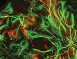
Supportive validation
- Submitted by
- Invitrogen Antibodies (provider)
- Main image

- Experimental details
- Immunocytochemistry analysis of alpha Internexin in Hippocampal neurons in tissue culture. Samples were incubated in alpha Internexin monoclonal antibody (Product # MA3-16726). Neurofilament alpha internexin (red), using alpha-Internexin Antibody and GFAP antibody (green).

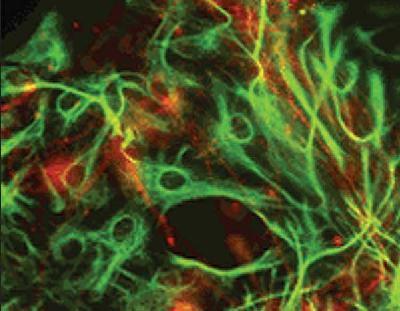
- Submitted by
- Invitrogen Antibodies (provider)
- Main image
- Experimental details
- Immunocytochemistry analysis of alpha Internexin in Hippocampal neurons in tissue culture. Samples were incubated in alpha Internexin monoclonal antibody (Product # MA3-16726). Neurofilament alpha internexin (red), using alpha-Internexin Antibody and GFAP antibody (green).
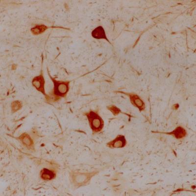
Supportive validation
- Submitted by
- Invitrogen Antibodies (provider)
- Main image
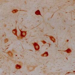
- Experimental details
- Immunohistochemical analysis of alpha Internexin in a section of rat facial nucleus 7 days following axotomy. Samples were incubated in alpha Internexin monoclonal antibody (Product # MA3-16726). These neurons are capable of regenerating their axons and also, concomitant with regeneration, strongly upregulate alpha-Internexin in their perikarya. Other central neurons which are not able to regenerate their axons do not upregulate this protein after axotomy and untreated facial neurons normally show only very low levels of alpha-Internexin. Both findings suggest that alpha-Internexin has a role in axonal regeneration.