 Explore
 Validate
 Learn
Learn Western blot
 Immunoprecipitation
Antibody data
- Antibody Data
- Antigen structure
- References [0]
- Comments [0]
- Validations
- Western blot [4]
- Immunocytochemistry [3]
- Immunohistochemistry [1]
Submit
Validation data
Reference
Comment
Report error
- Product number
- PA3-16725 - Provider product page

- Provider
- Invitrogen Antibodies
- Product name
- alpha Internexin Polyclonal Antibody
- Antibody type
- Polyclonal
- Antigen
- Purifed from natural sources
- Reactivity
- Human, Mouse, Rat, Bovine, Porcine
- Host
- Rabbit
- Isotype
- IgG
- Vial size
- 100 µL
- Concentration
- Conc. Not Determined
- Storage
- Store at 4°C short term. For long term storage, store at -20°C, avoiding freeze/thaw cycles.
No comments: Submit comment
Supportive validation
- Submitted by
- Invitrogen Antibodies (provider)
- Main image

- Experimental details
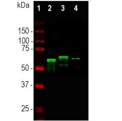
- Western blot analysis of alpha Internexin in whole tissue lysates. Samples were incubated in alpha Internexin polyclonal antibody (Product # PA3-16725 using a dilution of 1:10,000. Antibody in green: [1] protein standard (red), [2] mouse spinal cord, [3] rat spinal cord, [4] bovine spinal cord. Major bands in the 64-66 kDa range corresponds to alpha-internexin. The alpha-internexin protein from different species is known to vary slightly in SDS-PAGE molecular weight.

- Submitted by
- Invitrogen Antibodies (provider)
- Main image

- Experimental details
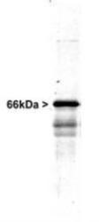
- Western blot analysis of alpha Internexin in whole rat spinal cord homogenate. Samples were incubated in alpha Internexin polyclonal antibody (Product # PA3-16725 using a dilution of 1:20,000. A prominent band running at ~66 kDa is apparent, as well as smaller lower bands which are apparently degradation products. A minor band at ~150 kDa is also seen, apparently resulting from dimerization of alpha-internexin.

- Submitted by
- Invitrogen Antibodies (provider)
- Main image
- Experimental details
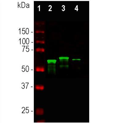
- Western blot analysis of alpha Internexin in whole tissue lysates. Samples were incubated in alpha Internexin polyclonal antibody (Product # PA3-16725 using a dilution of 1:10,000. Antibody in green: [1] protein standard (red), [2] mouse spinal cord, [3] rat spinal cord, [4] bovine spinal cord. Major bands in the 64-66 kDa range corresponds to alpha-internexin. The alpha-internexin protein from different species is known to vary slightly in SDS-PAGE molecular weight.
- Submitted by
- Invitrogen Antibodies (provider)
- Main image
- Experimental details
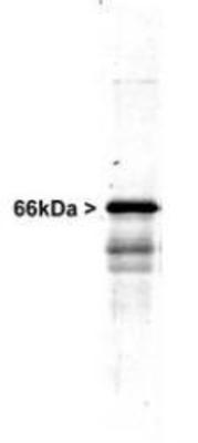
- Western blot analysis of alpha Internexin in whole rat spinal cord homogenate. Samples were incubated in alpha Internexin polyclonal antibody (Product # PA3-16725 using a dilution of 1:20,000. A prominent band running at ~66 kDa is apparent, as well as smaller lower bands which are apparently degradation products. A minor band at ~150 kDa is also seen, apparently resulting from dimerization of alpha-internexin.
Supportive validation
- Submitted by
- Invitrogen Antibodies (provider)
- Main image
- Experimental details
- Immunocytochemistry analysis of alpha Internexin in mixed neuron-glial cultures. Samples were incubated in alpha Internexin polyclonal antibody (Product # PA3-16725). Alpha-internexin (red) and chicken antibody to peripherin CPCA-Peri (green). The alpha internexin antibody stains numerous axonal and dendritic profiles in these cultures, while peripherin antibody binds to only a subset of neurons.
- Submitted by
- Invitrogen Antibodies (provider)
- Main image

- Experimental details
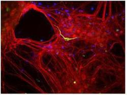
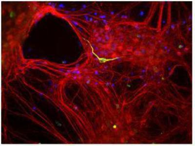
- Immunocytochemistry analysis of alpha Internexin in SHSY5Y cells. Samples were incubated in alpha Internexin polyclonal antibody (Product # PA3-16725) followed by Alexa Fluor 488-conjugated Goat to rabbit IgG secondary antibody (green). Actin filaments were labeled with Alexa Fluor 568 phalloidin (red). DAPI was used to stain the cell nuclei (blue).

- Submitted by
- Invitrogen Antibodies (provider)
- Main image

- Experimental details
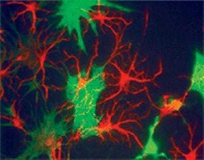
- Immunocytochemistry analysis of alpha Internexin in neuronal progenitor cells. Samples were incubated in alpha Internexin polyclonal antibody (Product # PA3-16725). Alpha internexin (red). The green stain shows the fibroblast marker, Plectin.
Supportive validation
- Submitted by
- Invitrogen Antibodies (provider)
- Main image
- Experimental details
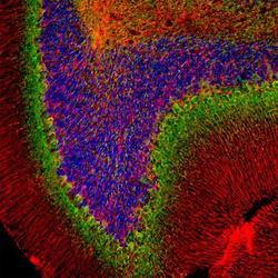
- Immunohistochemical analysis of alpha Internexin in Rat cerebellum section. Samples were incubated in alpha Internexin polyclonal antibody (Product # PA3-16725) using a dilution of 1:2000. Antibody in green, and chicken pAb to GFAP, dilution 1:5,000, in red. Blue is DAPI staining of nuclear DNA. Following transcardial perfusion with 4% paraformaldehyde, brain was post fixed for 24 hours, cut to 45 µM, and free-floating sections were stained with above antibodies. The alpha-internexin antibody selectively stains axons and dendrites of neuronal cells, in particular Purkinje cells and parallel fibers the axons of granule cells. The GFAP antibody labels network of glial cells, such as astrocytes in the granule cell layer and white matter and Bergmann glia in the molecular layer.
