 Explore
 Validate
 Learn
Learn Western blot
 Immunocytochemistry
Antibody data
- Antibody Data
- Antigen structure
- References [8]
- Comments [0]
- Validations
- Western blot [2]
- Immunohistochemistry [1]
Submit
Validation data
Reference
Comment
Report error
- Product number
- NB300-139 - Provider product page

- Provider
- Novus Biologicals
- Proper citation
- Novus Cat#NB300-139, RRID:AB_10001006
- Product name
- Rabbit Polyclonal alpha-Internexin Antibody
- Antibody type
- Polyclonal
- Description
- Unpurified.
- Reactivity
- Human, Mouse, Rat, Bovine, Porcine
- Host
- Rabbit
- Isotype
- IgG
- Vial size
- 0.1 ml
- Storage
- Store at 4C short term. Aliquot and store at -20C long term. Avoid freeze-thaw cycles.
Submitted references Congenital Anophthalmia and Binocular Neonatal Enucleation Differently Affect the Proteome of Primary and Secondary Visual Cortices in Mice.
Cortical murine neurons lacking the neurofilament light chain protein have an attenuated response to injury in vitro.
Cytoskeletal changes during development and aging in the cortex of neurofilament light protein knockout mice.
Degeneration of axons in spinal white matter in G93A mSOD1 mouse characterized by NFL and α-internexin immunoreactivity.
Neuron-glia interactions underlie ALS-like axonal cytoskeletal pathology.
Focal damage to the adult rat neocortex induces wound healing accompanied by axonal sprouting and dendritic structural plasticity.
Excitotoxicity mediated by non-NMDA receptors causes distal axonopathy in long-term cultured spinal motor neurons.
Investigation of general and cytoskeletal markers to estimate numbers and proportions of neurons in the human intestine.
Laramée ME, Smolders K, Hu TT, Bronchti G, Boire D, Arckens L
PloS one 2016;11(7):e0159320
PloS one 2016;11(7):e0159320
Cortical murine neurons lacking the neurofilament light chain protein have an attenuated response to injury in vitro.
Blizzard CA, King AE, Vickers J, Dickson T
Journal of neurotrauma 2013 Nov 15;30(22):1908-18
Journal of neurotrauma 2013 Nov 15;30(22):1908-18
Cytoskeletal changes during development and aging in the cortex of neurofilament light protein knockout mice.
Liu Y, Staal JA, Canty AJ, Kirkcaldie MT, King AE, Bibari O, Mitew ST, Dickson TC, Vickers JC
The Journal of comparative neurology 2013 Jun 1;521(8):1817-27
The Journal of comparative neurology 2013 Jun 1;521(8):1817-27
Degeneration of axons in spinal white matter in G93A mSOD1 mouse characterized by NFL and α-internexin immunoreactivity.
King AE, Blizzard CA, Southam KA, Vickers JC, Dickson TC
Brain research 2012 Jul 17;1465:90-100
Brain research 2012 Jul 17;1465:90-100
Neuron-glia interactions underlie ALS-like axonal cytoskeletal pathology.
King AE, Dickson TC, Blizzard CA, Woodhouse A, Foster SS, Chung RS, Vickers JC
Neurobiology of aging 2011 Mar;32(3):459-69
Neurobiology of aging 2011 Mar;32(3):459-69
Focal damage to the adult rat neocortex induces wound healing accompanied by axonal sprouting and dendritic structural plasticity.
Blizzard CA, Chuckowree JA, King AE, Hosie KA, McCormack GH, Chapman JA, Vickers JC, Dickson TC
Cerebral cortex (New York, N.Y. : 1991) 2011 Feb;21(2):281-91
Cerebral cortex (New York, N.Y. : 1991) 2011 Feb;21(2):281-91
Excitotoxicity mediated by non-NMDA receptors causes distal axonopathy in long-term cultured spinal motor neurons.
King AE, Dickson TC, Blizzard CA, Foster SS, Chung RS, West AK, Chuah MI, Vickers JC
The European journal of neuroscience 2007 Oct;26(8):2151-9
The European journal of neuroscience 2007 Oct;26(8):2151-9
Investigation of general and cytoskeletal markers to estimate numbers and proportions of neurons in the human intestine.
Ganns D, Schrödl F, Neuhuber W, Brehmer A
Histology and histopathology 2006 Jan;21(1):41-51
Histology and histopathology 2006 Jan;21(1):41-51
No comments: Submit comment
Supportive validation
- Submitted by
- Novus Biologicals (provider)
- Main image
- Experimental details
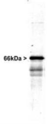
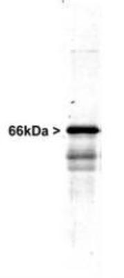
- Western Blot: alpha Internexin Antibody [NB300-139] - Western blot of whole rat spinal cord homogenate stained with RPCA-a-Int, at dilution of 1:20,000. A prominent band running at ~66kDa is apparent, as well as smaller lower bands which are apparently degradation products. A minor band at ~150kDa is also seen, apparently resulting from dimerization of alpha-internexin.
- Submitted by
- Novus Biologicals (provider)
- Main image
- Experimental details
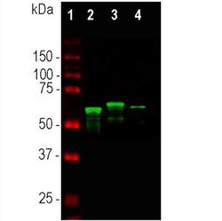
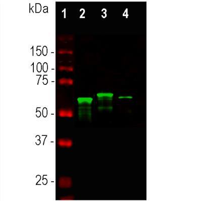
- Western Blot: alpha-Internexin Antibody [NB300-139] - Whole tissue lysates using rabbit pAb to alpha-internexin, dilution 1:10,000 in green: [1] protein standard (red), [2] mouse spinal cord, [3] rat spinal cord, [4] bovine spinal cord. Major bands in the 64-66 kDa range corresponds to alpha-internexin. The alpha-internexin protein from different species is known to vary slightly in SDS-PAGE molecular weight.
Supportive validation
- Submitted by
- Novus Biologicals (provider)
- Main image
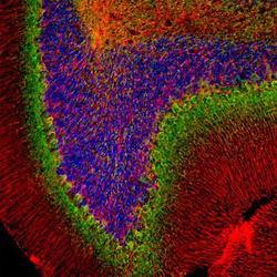
- Experimental details
- Immunohistochemistry: alpha-Internexin Antibody [NB300-139] - Rat cerebellum section stained with rabbit pAb to alpha-internexin, dilution 1:2,000, in green, and chicken pAb to GFAP, dilution 1:5,000, in red. Blue is DAPI staining of nuclear DNA. Following transcardial perfusion with 4% paraformaldehyde, brain was post fixed for 24 hours, cut to 45uM, and free-floating sections were stained with above antibodies. The alpha-internexin antibody selectively stains axons and dendrites of neuronal cells, in particular Purkinje cells and parallel fibers the axons of granule cells. The GFAP antibody labels network of glial cells, such as astrocytes in the granule cell layer and white matter and Bergmann glia in the molecular layer.
