Explore
 Validate
 Learn
Learn Western blot
 Immunocytochemistry
Antibody data
- Antibody Data
- Antigen structure
- References [1]
- Comments [0]
- Validations
- Western blot [1]
- Other assay [1]
Submit
Validation data
Reference
Comment
Report error
- Product number
- NB300-216 - Provider product page

- Provider
- Novus Biologicals
- Proper citation
- Novus Cat#NB300-216, RRID:AB_10081414
- Product name
- Mouse Monoclonal alpha-Internexin Antibody
- Antibody type
- Monoclonal
- Antigen sequence
The epitope for this antibody is no
t in the N-terminus
Submitted references Angiotensin II increases GABAB receptor expression in nucleus tractus solitarii of rats.
Yao F, Sumners C, O'Rourke ST, Sun C
American journal of physiology. Heart and circulatory physiology 2008 Jun;294(6):H2712-20
American journal of physiology. Heart and circulatory physiology 2008 Jun;294(6):H2712-20
No comments: Submit comment
Supportive validation
- Submitted by
- Novus Biologicals (provider)
- Main image
- Experimental details
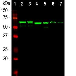
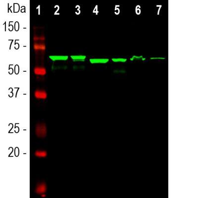
- Western Blot: alpha-Internexin Antibody (1D2) [NB300-216] - Analysis of different tissue lysates using alpha-Internexin antibody, dilution 1:10,000 (Green): [1] protein standard, [2] rat brain, [3] rat spinal cord, [4] mouse brain, [5] mouse spinal cord, [6] pig spinal cord and [7] cow spinal cord. The alpha-Internexin antibody reveals the alpha-internexin protein with apparent molecular weight of 64 to 66 kDa with slight variability among species.
Supportive validation
- Submitted by
- Novus Biologicals (provider)
- Main image

- Experimental details
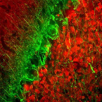
- Immunohistochemistry Free-Floating: alpha-Internexin Antibody (1D2) [NB300-216] - Analysis of rat cerebellum section stained with alpha-internexin antibody, dilution 1:5,000 (Green), and costained with chicken Calretinin pAb, dilution 1:2,000 (Red). Following transcardial perfusion of rat with 4% paraformaldehyde, brain was post fixed for 24hrs, cut to 45uM, and free-floating sections were stained with the above antibodies. The alpha-internexin antibody selectively stains neuronal processes, in particular parallel fibers, the axons of granule cells. Calretinin antibody stains interneurons predominantly in the molecular layer of the cerebellum.