Explore
 Validate
 Learn
Learn Western blot
 Immunohistochemistry
Antibody data
- Antibody Data
- Antigen structure
- References [7]
- Comments [0]
- Validations
- Immunohistochemistry [1]
Submit
Validation data
Reference
Comment
Report error
- Product number
- HPA018139 - Provider product page

- Provider
- Atlas Antibodies
- Proper citation
- Atlas Antibodies Cat#HPA018139, RRID:AB_1849903
- Product name
- Anti-GOT2
- Antibody type
- Polyclonal
- Description
- Polyclonal Antibody against Human GOT2, Gene description: glutamic-oxaloacetic transaminase 2, mitochondrial, Alternative Gene Names: KAT4, KATIV, mitAAT, Validated applications: IHC, WB, Uniprot ID: P00505, Storage: Store at +4°C for short term storage. Long time storage is recommended at -20°C.
- Reactivity
- Human, Mouse, Rat
- Host
- Rabbit
- Conjugate
- Unconjugated
- Isotype
- IgG
- Vial size
- 100 µl
- Concentration
- 0.1 mg/ml
- Storage
- Store at +4°C for short term storage. Long time storage is recommended at -20°C.
- Handling
- The antibody solution should be gently mixed before use.
Submitted references HIF1α-dependent uncoupling of glycolysis suppresses tumor cell proliferation
Loss of mitochondrial enzyme GPT2 leads to reprogramming of synaptic glutamate metabolism
Requirement of hepatic pyruvate carboxylase during fasting, high fat, and ketogenic diet
A Cancer Cell–Intrinsic GOT2–PPARδ Axis Suppresses Antitumor Immunity
Metabolic requirement for GOT2 in pancreatic cancer depends on environmental context
Interplay Between Thiamine and p53/p21 Axes Affects Antiproliferative Action of Cisplatin in Lung Adenocarcinoma Cells by Changing Metabolism of 2-Oxoglutarate/Glutamate
Tissue of origin dictates GOT1 dependence and confers synthetic lethality to radiotherapy.
Urrutia A, Mesa-Ciller C, Guajardo-Grence A, Alkan H, Soro-Arnáiz I, Vandekeere A, Ferreira Campos A, Igelmann S, Fernández-Arroyo L, Rinaldi G, Lorendeau D, De Bock K, Fendt S, Aragonés J
Cell Reports 2024;43(4):114103
Cell Reports 2024;43(4):114103
Loss of mitochondrial enzyme GPT2 leads to reprogramming of synaptic glutamate metabolism
Baytas O, Davidson S, Kauer J, Morrow E
Molecular Brain 2024;17(1)
Molecular Brain 2024;17(1)
Requirement of hepatic pyruvate carboxylase during fasting, high fat, and ketogenic diet
Selen E, Rodriguez S, Cavagnini K, Kim H, Na C, Wolfgang M
Journal of Biological Chemistry 2022;298(12):102648
Journal of Biological Chemistry 2022;298(12):102648
A Cancer Cell–Intrinsic GOT2–PPARδ Axis Suppresses Antitumor Immunity
Abrego J, Sanford-Crane H, Oon C, Xiao X, Betts C, Sun D, Nagarajan S, Diaz L, Sandborg H, Bhattacharyya S, Xia Z, Coussens L, Tontonoz P, Sherman M
Cancer Discovery 2022;12(10):2414-2433
Cancer Discovery 2022;12(10):2414-2433
Metabolic requirement for GOT2 in pancreatic cancer depends on environmental context
Kerk S, Lin L, Myers A, Sutton D, Andren A, Sajjakulnukit P, Zhang L, Zhang Y, Jiménez J, Nelson B, Chen B, Robinson A, Thurston G, Kemp S, Steele N, Hoffman M, Wen H, Long D, Ackenhusen S, Ramos J, Gao X, Nwosu Z, Galban S, Halbrook C, Lombard D, Piwnica-Worms D, Ying H, Pasca di Magliano M, Crawford H, Shah Y, Lyssiotis C
eLife 2022;11
eLife 2022;11
Interplay Between Thiamine and p53/p21 Axes Affects Antiproliferative Action of Cisplatin in Lung Adenocarcinoma Cells by Changing Metabolism of 2-Oxoglutarate/Glutamate
Aleshin V, Zhou X, Krishnan S, Karlsson A, Bunik V
Frontiers in Genetics 2021;12
Frontiers in Genetics 2021;12
Tissue of origin dictates GOT1 dependence and confers synthetic lethality to radiotherapy.
Nelson BS, Lin L, Kremer DM, Sousa CM, Cotta-Ramusino C, Myers A, Ramos J, Gao T, Kovalenko I, Wilder-Romans K, Dresser J, Davis M, Lee HJ, Nwosu ZC, Campit S, Mashadova O, Nicolay BN, Tolstyka ZP, Halbrook CJ, Chandrasekaran S, Asara JM, Crawford HC, Cantley LC, Kimmelman AC, Wahl DR, Lyssiotis CA
Cancer & metabolism 2020;8:1
Cancer & metabolism 2020;8:1
No comments: Submit comment
Supportive validation
- Submitted by
- Atlas Antibodies (provider)
- Enhanced method
- Orthogonal validation
- Main image
- Experimental details
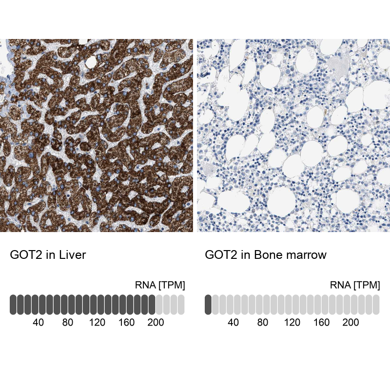
- Immunohistochemistry analysis in human liver and bone marrow tissues using HPA018139 antibody. Corresponding GOT2 RNA-seq data are presented for the same tissues.
- Sample type
- Human
- Protocol
- Protocol