Explore
 Validate
 Learn
Learn Western blot
Antibody data
- Antibody Data
- Antigen structure
- References [4]
- Comments [0]
- Validations
- Western blot [2]
- Immunohistochemistry [1]
Submit
Validation data
Reference
Comment
Report error
- Product number
- AF1048 - Provider product page

- Provider
- R&D Systems
- Product name
- Human HAI-1 Antibody
- Antibody type
- Polyclonal
- Description
- Antigen Affinity-purified. Detects human HAI-1 in direct ELISAs and Western blots. In direct ELISAs, approximately 20% cross-reactivity with recombinant mouse HAI-1 is observed and less than 1% cross-reactivity with recombinant human HAI-2 is observed.
- Reactivity
- Human
- Host
- Goat
- Conjugate
- Unconjugated
- Antigen sequence
NP_003701- Isotype
- IgG
- Vial size
- 100 ug
- Concentration
- LYOPH
- Storage
- Use a manual defrost freezer and avoid repeated freeze-thaw cycles. 12 months from date of receipt, -20 to -70 °C as supplied. 1 month, 2 to 8 °C under sterile conditions after reconstitution. 6 months, -20 to -70 °C under sterile conditions after reconstitution.
Submitted references Phosphorylation of the type II transmembrane serine protease, TMPRSS13, in hepatocyte growth factor activator inhibitor-1 and -2-mediated cell-surface localization.
Matrix metalloproteinase-7 induces homotypic tumor cell aggregation via proteolytic cleavage of the membrane-bound Kunitz-type inhibitor HAI-1.
Prostasin is required for matriptase activation in intestinal epithelial cells to regulate closure of the paracellular pathway.
Matriptase inhibition by hepatocyte growth factor activator inhibitor-1 is essential for placental development.
Murray AS, Varela FA, Hyland TE, Schoenbeck AJ, White JM, Tanabe LM, Todi SV, List K
The Journal of biological chemistry 2017 Sep 8;292(36):14867-14884
The Journal of biological chemistry 2017 Sep 8;292(36):14867-14884
Matrix metalloproteinase-7 induces homotypic tumor cell aggregation via proteolytic cleavage of the membrane-bound Kunitz-type inhibitor HAI-1.
Ishikawa T, Kimura Y, Hirano H, Higashi S
The Journal of biological chemistry 2017 Dec 15;292(50):20769-20784
The Journal of biological chemistry 2017 Dec 15;292(50):20769-20784
Prostasin is required for matriptase activation in intestinal epithelial cells to regulate closure of the paracellular pathway.
Buzza MS, Martin EW, Driesbaugh KH, Désilets A, Leduc R, Antalis TM
The Journal of biological chemistry 2013 Apr 12;288(15):10328-37
The Journal of biological chemistry 2013 Apr 12;288(15):10328-37
Matriptase inhibition by hepatocyte growth factor activator inhibitor-1 is essential for placental development.
Szabo R, Molinolo A, List K, Bugge TH
Oncogene 2007 Mar 8;26(11):1546-56
Oncogene 2007 Mar 8;26(11):1546-56
No comments: Submit comment
Supportive validation
- Submitted by
- R&D Systems (provider)
- Main image

- Experimental details
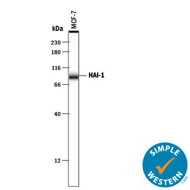
- Detection of Human HAI-1 by Simple WesternTM. Simple Western lane view shows lysates of MCF-7 human breast cancer cell line, loaded at 0.2 mg/mL. A specific band was detected for HAI-1 at approximately 88 kDa (as indicated) using 10 µg/mL of Goat Anti-Human HAI-1 Antigen Affinity-purified Polyclonal Antibody (Catalog # AF1048) followed by 1:50 dilution of HRP-conjugated Anti-Goat IgG Secondary Antibody (Catalog # HAF109). This experiment was conducted under reducing conditions and using the 12-230 kDa separation system. Non-specific interaction with the 230 kDa Simple Western standard may be seen with this antibody.
- Submitted by
- R&D Systems (provider)
- Main image
- Experimental details
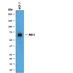
- Detection of Human HAI-1 by Western Blot. Western blot shows lysates of MCF-7 human breast cancer cell line. PVDF membrane was probed with 1 µg/mL of Goat Anti-Human HAI-1 Antigen Affinity-purified Polyclonal Antibody (Catalog # AF1048) followed by HRP-conjugated Anti-Goat IgG Secondary Antibody (Catalog # HAF019). A specific band was detected for HAI-1 at approximately 70 kDa (as indicated). This experiment was conducted under reducing conditions and using Immunoblot Buffer Group 1.

Supportive validation
- Submitted by
- R&D Systems (provider)
- Main image
- Experimental details
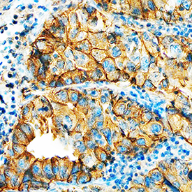
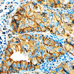
- HAI-1 in Human Lung Cancer Tissue. HAI-1 was detected in immersion fixed paraffin-embedded sections of human lung cancer tissue using Goat Anti-Human HAI-1 Antigen Affinity-purified Polyclonal Antibody (Catalog # AF1048) at 5 µg/mL overnight at 4 °C. Tissue was stained using the Anti-Goat HRP-DAB Cell & Tissue Staining Kit (brown; Catalog # CTS008) and counterstained with hematoxylin (blue). Specific staining was localized to plasma membrane in epithelial cells. View our protocol for Chromogenic IHC Staining of Paraffin-embedded Tissue Sections.