 Explore
 Validate
 Learn
Learn Western blot
 Immunohistochemistry
Antibody data
- Antibody Data
- Antigen structure
- References [7]
- Comments [0]
- Validations
- Immunohistochemistry [2]
- Flow cytometry [3]
Submit
Validation data
Reference
Comment
Report error
- Product number
- NB300-539 - Provider product page

- Provider
- Novus Biologicals
- Proper citation
- Novus Cat#NB300-539, RRID:AB_2085316
- Product name
- Mouse Monoclonal CRABP1 Antibody
- Antibody type
- Monoclonal
- Description
- Unpurified. Detects cellular Retinoic Acid Binding Protein 1 (CRABP 1). This does not cross-react with Cellular Retinol Binding Protein (CRBP), performic acid oxidized CRBP, interphotoreceptor retinoid binding protein or retinol binding protein.
- Reactivity
- Human, Mouse, Rat, Bovine, Canine, Chicken/Avian, Feline, Rabbit, Simian
- Host
- Mouse
- Isotype
- IgG
- Vial size
- 100uL
- Concentration
- 5 mg/ml
- Storage
- Store at -20C. Avoid freeze-thaw cycles.
Submitted references Retinoic acid promotes differentiation of photoreceptors in vitro.
The developing organ of Corti contains retinoic acid and forms supernumerary hair cells in response to exogenous retinoic acid in culture.
Localization of cellular retinoic acid-binding protein to amacrine cells of rat retina.
Localization of cellular retinoic acid-binding protein to amacrine cells of rat retina.
Immunolocalization of cellular retinoic acid binding protein to Müller cells and/or a subpopulation of GABA-positive amacrine cells in retinas of different species.
Immunolocalization of cellular retinol-, retinaldehyde- and retinoic acid-binding proteins in rat retina during pre- and postnatal development.
Immunolocalization of cellular retinol-, retinaldehyde- and retinoic acid-binding proteins in rat retina during pre- and postnatal development.
Kelley MW, Turner JK, Reh TA
Development (Cambridge, England) 1994 Aug;120(8):2091-102
Development (Cambridge, England) 1994 Aug;120(8):2091-102
The developing organ of Corti contains retinoic acid and forms supernumerary hair cells in response to exogenous retinoic acid in culture.
Kelley MW, Xu XM, Wagner MA, Warchol ME, Corwin JT
Development (Cambridge, England) 1993 Dec;119(4):1041-53
Development (Cambridge, England) 1993 Dec;119(4):1041-53
Localization of cellular retinoic acid-binding protein to amacrine cells of rat retina.
Gaur VP, de Leeuw AM, Milam AH, Saari JC
Experimental eye research 1990 May;50(5):505-11
Experimental eye research 1990 May;50(5):505-11
Localization of cellular retinoic acid-binding protein to amacrine cells of rat retina.
Gaur VP, de Leeuw AM, Milam AH, Saari JC
Experimental eye research 1990 May;50(5):505-11
Experimental eye research 1990 May;50(5):505-11
Immunolocalization of cellular retinoic acid binding protein to Müller cells and/or a subpopulation of GABA-positive amacrine cells in retinas of different species.
Milam AH, De Leeuw AM, Gaur VP, Saari JC
The Journal of comparative neurology 1990 Jun 1;296(1):123-9
The Journal of comparative neurology 1990 Jun 1;296(1):123-9
Immunolocalization of cellular retinol-, retinaldehyde- and retinoic acid-binding proteins in rat retina during pre- and postnatal development.
De Leeuw AM, Gaur VP, Saari JC, Milam AH
Journal of neurocytology 1990 Apr;19(2):253-64
Journal of neurocytology 1990 Apr;19(2):253-64
Immunolocalization of cellular retinol-, retinaldehyde- and retinoic acid-binding proteins in rat retina during pre- and postnatal development.
De Leeuw AM, Gaur VP, Saari JC, Milam AH
Journal of neurocytology 1990 Apr;19(2):253-64
Journal of neurocytology 1990 Apr;19(2):253-64
No comments: Submit comment
Supportive validation
- Submitted by
- Novus Biologicals (provider)
- Main image
- Experimental details
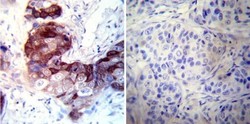
- Immunohistochemistry-Paraffin: CRABP1 Antibody (C-1) [NB300-539] - Both normal and cancer biopsies of deparaffinized Human breast carcinoma tissues.

- Submitted by
- Novus Biologicals (provider)
- Main image
- Experimental details
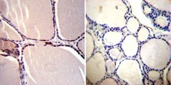
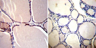
- Immunohistochemistry-Paraffin: CRABP1 Antibody (C-1) [NB300-539] - Both normal and cancer biopsies of deparaffinized Human thyroid tissues.
Supportive validation
- Submitted by
- Novus Biologicals (provider)
- Main image

- Experimental details
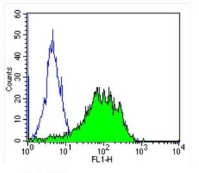
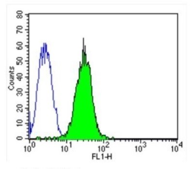
- Flow Cytometry: CRABP1 Antibody (C-1) [NB300-539] - Analysis of CRABPI in NIH-3T3 cells (green) compared to an isotype control (blue).
- Submitted by
- Novus Biologicals (provider)
- Main image

- Experimental details
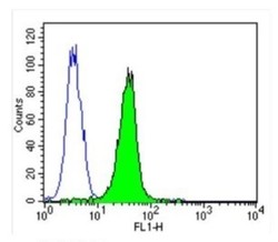
- Flow Cytometry: CRABP1 Antibody (C-1) [NB300-539] - Analysis of CRABPI in MCF-7 cells (green) compared to an isotype control (blue).
- Submitted by
- Novus Biologicals (provider)
- Main image
- Experimental details
- Flow Cytometry: CRABP1 Antibody (C-1) [NB300-539] - Analysis of CRABPI in Hela cells (green) compared to an isotype control (blue).
