 Explore
 Validate
 Learn
Learn Western blot
 Immunocytochemistry
Antibody data
- Antibody Data
- Antigen structure
- References [2]
- Comments [0]
- Validations
- Western blot [4]
- Immunohistochemistry [4]
Submit
Validation data
Reference
Comment
Report error
- Product number
- NBP1-32244 - Provider product page

- Provider
- Novus Biologicals
- Proper citation
- Novus Cat#NBP1-32244, RRID:AB_10003923
- Product name
- Rabbit Polyclonal Calretinin Antibody
- Antibody type
- Polyclonal
- Description
- Immunogen affinity purified.
- Reactivity
- Human, Mouse, Rat
- Host
- Rabbit
- Isotype
- IgG
- Vial size
- 0.1 ml
- Storage
- Aliquot and store at -20C or -80C. Avoid freeze-thaw cycles.
Submitted references Dual embryonic origin of the mammalian enteric nervous system.
Male germ cells support long-term propagation of Zika virus.
Brokhman I, Xu J, Coles BLK, Razavi R, Engert S, Lickert H, Babona-Pilipos R, Morshead CM, Sibley E, Chen C, van der Kooy D
Developmental biology 2019 Jan 15;445(2):256-270
Developmental biology 2019 Jan 15;445(2):256-270
Male germ cells support long-term propagation of Zika virus.
Robinson CL, Chong ACN, Ashbrook AW, Jeng G, Jin J, Chen H, Tang EI, Martin LA, Kim RS, Kenyon RM, Do E, Luna JM, Saeed M, Zeltser L, Ralph H, Dudley VL, Goldstein M, Rice CM, Cheng CY, Seandel M, Chen S
Nature communications 2018 May 29;9(1):2090
Nature communications 2018 May 29;9(1):2090
No comments: Submit comment
Supportive validation
- Submitted by
- Novus Biologicals (provider)
- Main image

- Experimental details
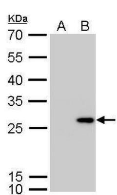
- Western Blot: Calretinin Antibody [NBP1-32244] - A. 30 ug 293T whole cell lysate/extract. B. 30 ug whole cell lysate/extract of human CALB2-transfected 293T cells.
- Submitted by
- Novus Biologicals (provider)
- Main image
- Experimental details
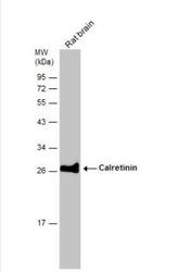
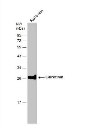
- Western Blot: Calretinin Antibody [NBP1-32244] - Rat tissue extract (50 ug) was separated by 12% SDS-PAGE, and the membrane was blotted with Calretinin antibody diluted at 1:1000. The HRP-conjugated anti-rabbit IgG antibody (NBP2-19301) was used to detect the primary antibody.
- Submitted by
- Novus Biologicals (provider)
- Main image

- Experimental details
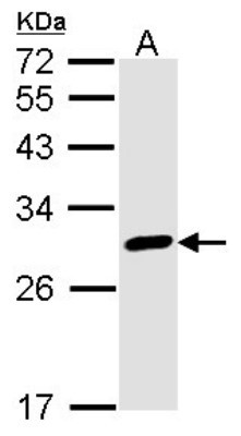
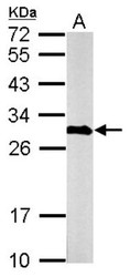
- Western Blot: Calretinin Antibody [NBP1-32244] - Sample (30 ug of whole cell lysate) A: A431 12% SDS PAGE Calretinin antibody, antibody diluted at 1:1000.
- Submitted by
- Novus Biologicals (provider)
- Main image
- Experimental details
- Western Blot: Calretinin Antibody [NBP1-32244] - Sample (50 ug of whole cell lysate) A: mouse brain 12% SDS PAGE; antibody diluted at 1:1000.

Supportive validation
- Submitted by
- Novus Biologicals (provider)
- Main image
- Experimental details
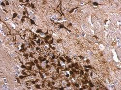
- Immunohistochemistry-Paraffin: Calretinin Antibody [NBP1-32244] - Paraffin-embedded mouse fore brain. Calretinin antibody dilution: 1:500.

- Submitted by
- Novus Biologicals (provider)
- Main image
- Experimental details
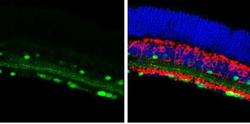
- Immunohistochemistry-Frozen: Calretinin Antibody [NBP1-32244] - Frozen sectioned adult mouse retina. Green: Calretinin protein stained by Calretinin antibody diluted at 1:250. Red: Protein kinase C alpha staining. Blue: Fluoroshield with DAPI.

- Submitted by
- Novus Biologicals (provider)
- Main image

- Experimental details
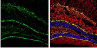
- Immunohistochemistry-Frozen: Calretinin Antibody [NBP1-32244] - Frozen Section adult mouse hippocampus. Green: Calretinin protein stained by Calretinin antibody diluted at 1:250. Red: NF-H, stained by NF-H antibody [114] diluted at 1:500. Blue: Fluoroshield with DAPI.
- Submitted by
- Novus Biologicals (provider)
- Main image
- Experimental details
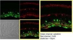
- Immunohistochemistry-Paraffin: Calretinin Antibody [NBP1-32244] - Analysis of paraffin-embedded Mouse retina, using Calretinin antibody at 1:250 dilution.
