 Explore
 Validate
 Learn
Learn Western blot
Antibody data
- Antibody Data
- Antigen structure
- References [3]
- Comments [0]
- Validations
- Western blot [1]
- Immunohistochemistry [1]
Submit
Validation data
Reference
Comment
Report error
- Product number
- AF5065 - Provider product page

- Provider
- R&D Systems
- Product name
- Human/Mouse/Rat Calretinin Antibody
- Antibody type
- Polyclonal
- Description
- Immunogen affinity purified. Detects human, mouse, and rat Calretinin in direct ELISAs and Western blots. In direct ELISAs, this antibody shows approximately 5% cross-reactivity with recombinant human Calbindin D.
- Reactivity
- Human, Mouse, Rat
- Host
- Goat
- Conjugate
- Unconjugated
- Antigen sequence
AAH15484- Isotype
- IgG
- Vial size
- 100 ug
- Concentration
- LYOPH
- Storage
- Use a manual defrost freezer and avoid repeated freeze-thaw cycles. 12 months from date of receipt, -20 to -70 °C as supplied. 1 month, 2 to 8 °C under sterile conditions after reconstitution. 6 months, -20 to -70 °C under sterile conditions after reconstitution.
Submitted references The nucleus reuniens: a key node in the neurocircuitry of stress and depression.
Estrogen Treatment Reverses Prematurity-Induced Disruption in Cortical Interneuron Population.
AAV2-Mediated Transduction of the Mouse Retina After Optic Nerve Injury.
Kafetzopoulos V, Kokras N, Sotiropoulos I, Oliveira JF, Leite-Almeida H, Vasalou A, Sardinha VM, Papadopoulou-Daifoti Z, Almeida OFX, Antoniou K, Sousa N, Dalla C
Molecular psychiatry 2018 Mar;23(3):579-586
Molecular psychiatry 2018 Mar;23(3):579-586
Estrogen Treatment Reverses Prematurity-Induced Disruption in Cortical Interneuron Population.
Panda S, Dohare P, Jain S, Parikh N, Singla P, Mehdizadeh R, Klebe DW, Kleinman GM, Cheng B, Ballabh P
The Journal of neuroscience : the official journal of the Society for Neuroscience 2018 Aug 22;38(34):7378-7391
The Journal of neuroscience : the official journal of the Society for Neuroscience 2018 Aug 22;38(34):7378-7391
AAV2-Mediated Transduction of the Mouse Retina After Optic Nerve Injury.
Nickells RW, Schmitt HM, Maes ME, Schlamp CL
Investigative ophthalmology & visual science 2017 Dec 1;58(14):6091-6104
Investigative ophthalmology & visual science 2017 Dec 1;58(14):6091-6104
No comments: Submit comment
Supportive validation
- Submitted by
- R&D Systems (provider)
- Main image
- Experimental details
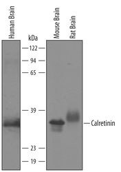
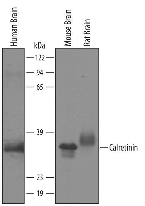
- Detection of Human/Mouse/Rat Calretinin by Western Blot. Western blot shows lysates of human brain, mouse brain and rat brain tissue. PVDF membrane was probed with 1 µg/mL of Goat Anti-Human/Mouse/Rat Calretinin Antigen Affinity-purified Polyclonal Antibody (Catalog # AF5065) followed by HRP-conjugated Anti-Goat IgG Secondary Antibody (Catalog # HAF019). A specific band was detected for Calretinin at approximately 31 kDa (as indicated). This experiment was conducted under reducing conditions and using Immunoblot Buffer Group 8.
Supportive validation
- Submitted by
- R&D Systems (provider)
- Main image
- Experimental details
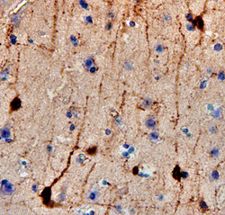
- Calretinin in Human Brain. Calretinin was detected in immersion fixed paraffin-embedded sections of human brain (cortex) using 15 µg/mL Goat Anti-Human/Mouse/ Rat Calretinin Antigen Affinity-purified Polyclonal Antibody (Catalog # AF5065) overnight at 4 °C. Tissue was stained with the Anti-Goat HRP-DAB Cell & Tissue Staining Kit (brown; Catalog # CTS008) and counterstained with hematoxylin (blue). View our protocol for Chromogenic IHC Staining of Paraffin-embedded Tissue Sections.
