 Explore
 Validate
 Learn
Learn Western blot
 Immunocytochemistry
Antibody data
- Antibody Data
- Antigen structure
- References [2]
- Comments [0]
- Validations
- Western blot [1]
- Immunocytochemistry [1]
- Immunohistochemistry [1]
Submit
Validation data
Reference
Comment
Report error
- Product number
- HPA007305 - Provider product page

- Provider
- Atlas Antibodies
- Proper citation
- Atlas Antibodies Cat#HPA007305, RRID:AB_1078386
- Product name
- Anti-CALB2
- Antibody type
- Polyclonal
- Description
- Polyclonal Antibody against Human CALB2, Gene description: calbindin 2, Alternative Gene Names: CAL2, Validated applications: ICC, IHC, WB, Uniprot ID: P22676, Storage: Store at +4°C for short term storage. Long time storage is recommended at -20°C.
- Reactivity
- Human
- Host
- Rabbit
- Conjugate
- Unconjugated
- Isotype
- IgG
- Vial size
- 100 µl
- Concentration
- 0.6 mg/ml
- Storage
- Store at +4°C for short term storage. Long time storage is recommended at -20°C.
- Handling
- The antibody solution should be gently mixed before use.
Submitted references A human multi-cellular model shows how platelets drive production of diseased extracellular matrix and tissue invasion
Tissue Profiling of the Mammalian Central Nervous System Using Human Antibody-based Proteomics
Malacrida B, Nichols S, Maniati E, Jones R, Delanie-Smith R, Roozitalab R, Tyler E, Thomas M, Boot G, Mackerodt J, Lockley M, Knight M, Balkwill F, Pearce O
iScience 2021;24(6):102676
iScience 2021;24(6):102676
Tissue Profiling of the Mammalian Central Nervous System Using Human Antibody-based Proteomics
Mulder J, Björling E, Jonasson K, Wernérus H, Hober S, Hökfelt T, Uhlén M
Molecular & Cellular Proteomics 2009;8(7):1612-1622
Molecular & Cellular Proteomics 2009;8(7):1612-1622
No comments: Submit comment
Enhanced validation
- Submitted by
- Atlas Antibodies (provider)
- Enhanced method
- Genetic validation
- Main image
- Experimental details
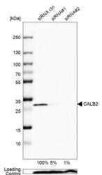
- Western blot analysis in U2OS cells transfected with control siRNA, target specific siRNA probe #1 and #2, using Anti-CALB2 antibody. Remaining relative intensity is presented. Loading control: Anti-PPIB.
- Sample type
- Human
- Protocol
- Protocol

Supportive validation
- Submitted by
- Atlas Antibodies (provider)
- Main image
- Experimental details
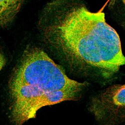
- Immunofluorescent staining of human cell line U-2 OS shows localization to cytosol.
- Sample type
- Human

Supportive validation
- Submitted by
- Atlas Antibodies (provider)
- Enhanced method
- Orthogonal validation
- Main image
- Experimental details
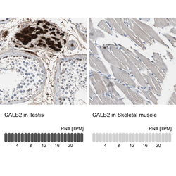
- Immunohistochemistry analysis in human testis and skeletal muscle tissues using HPA007305 antibody. Corresponding CALB2 RNA-seq data are presented for the same tissues.
- Sample type
- Human
- Protocol
- Protocol
