Explore
 Validate
 Learn
LearnPA5-26222
antibody from Invitrogen Antibodies
Targeting: ASIC2
ACCN, ACCN1, ASIC2a, BNaC1, BNC1, hBNaC1, MDEG
 Western blot
 Immunohistochemistry
Antibody data
- Antibody Data
- Antigen structure
- References [2]
- Comments [0]
- Validations
- Immunohistochemistry [1]
- Flow cytometry [2]
- Other assay [5]
Submit
Validation data
Reference
Comment
Report error
- Product number
- PA5-26222 - Provider product page

- Provider
- Invitrogen Antibodies
- Product name
- ASIC2 Polyclonal Antibody
- Antibody type
- Polyclonal
- Antigen
- Synthetic peptide
- Description
- This antibody is predicted to react with mouse and rat based on sequence homology.
- Reactivity
- Human
- Host
- Rabbit
- Isotype
- IgG
- Vial size
- 400 μL
- Concentration
- 0.3 mg/mL
- Storage
- Store at 4°C short term. For long term storage, store at -20°C, avoiding freeze/thaw cycles.
Submitted references Expression Profiles of ASIC1/2 and TRPV1/4 in Common Skin Tumors.
pH sensing in skin tumors: Methods to study the involvement of GPCRs, acid-sensing ion channels and transient receptor potential vanilloid channels.
Ackermann K, Wallner S, Brochhausen C, Schreml S
International journal of molecular sciences 2021 Jun 2;22(11)
International journal of molecular sciences 2021 Jun 2;22(11)
pH sensing in skin tumors: Methods to study the involvement of GPCRs, acid-sensing ion channels and transient receptor potential vanilloid channels.
Stolwijk JA, Sauer L, Ackermann K, Nassios A, Aung T, Haerteis S, Bäumner AJ, Wegener J, Schreml S
Experimental dermatology 2020 Nov;29(11):1055-1061
Experimental dermatology 2020 Nov;29(11):1055-1061
No comments: Submit comment
Supportive validation
- Submitted by
- Invitrogen Antibodies (provider)
- Main image
- Experimental details
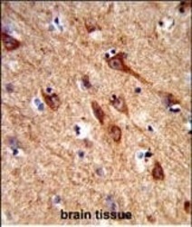
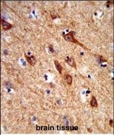
- Immunohistochemistry analysis of ASIC2 in formalin-fixed and paraffin-embedded human brain tissue. Samples were incubated with ASIC2 polyclonal antibody (Product # PA5-26222) which was peroxidase-conjugated to the secondary antibody, followed by DAB staining. This data demonstrates the use of this antibody for immunohistochemistry; clinical relevance has not been evaluated.
Supportive validation
- Submitted by
- Invitrogen Antibodies (provider)
- Main image

- Experimental details
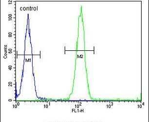
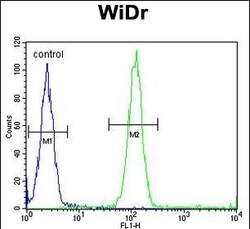
- Flow cytometry analysis of WiDr cells using an ACCN1 polyclonal antibody (Product # PA5-26222) (right) compared to a negative control cell (left) at a dilution of 1:10-50, followed by a FITC-conjugated goat anti-rabbit antibody
- Submitted by
- Invitrogen Antibodies (provider)
- Main image
- Experimental details
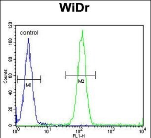
- Flow cytometry of ASIC2 in WiDr cells (right histogram). Samples were incubated with ASIC2 polyclonal antibody (Product # PA5-26222) followed by FITC-conjugated goat-anti-rabbit secondary antibody. Negative control cell (left histogram).
Supportive validation
- Submitted by
- Invitrogen Antibodies (provider)
- Main image
- Experimental details
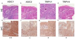
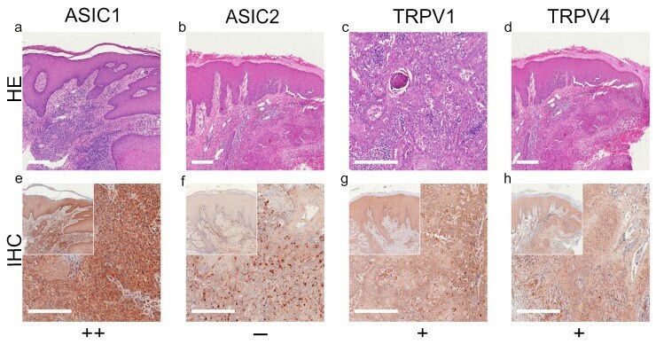
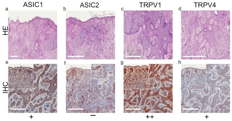
- Figure 1 Immunohistochemistry of SCC. Immunohistochemical staining for ASIC1, ASIC2, TRPV1 and TRPV4 in SCC tissue. ( a - d ) H&E staining, ( e - h ) immunohistochemical staining, inserted smaller pictures represent a two times larger perspective. Scale bars represent 200 mum. ( a - h ) Patient 8. This SCC shows no expression of ASIC2, only some peritumoral lymphocytes appear positive. The tumor cells show a weak, positive expression of TRPV1 and TRPV4. ASIC1 is expressed strongly on tumor cells. For more stainings of other SCCs, see Supplementary Figures S1-S3 .
- Submitted by
- Invitrogen Antibodies (provider)
- Main image

- Experimental details
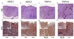
- Figure 2 Immunohistochemistry of BCC. Immunohistochemical staining for ASIC1, ASIC2, TRPV1 and TRPV4 in BCC tissue. ( a - d ) H&E staining, ( e - h ) immunohistochemical staining, inserted smaller pictures give an overview. Scale bars represent 200 mum. ( a - h ) Patient 10. This BCC shows a strong expression of ASIC1 and TRPV1. The expression of TRPV4 is weak and positive, but this BCC shows no expression of ASIC2. For more stainings of other BCCs, see Supplementary Figures S4-S7 .
- Submitted by
- Invitrogen Antibodies (provider)
- Main image
- Experimental details
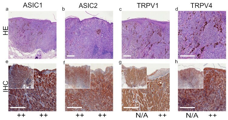
- Figure 4 Immunohistochemistry of MM. Immunohistochemical staining for ASIC1, ASIC2, TRPV1 and TRPV4 in MM tissue. ( a - d ) Histochemical H&E staining, ( e - h ) immunohistochemical staining, inserted smaller pictures represent a two times larger perspective. Scale bars represent 200 mum. ( a - h ) Patient 30.
- Submitted by
- Invitrogen Antibodies (provider)
- Main image

- Experimental details
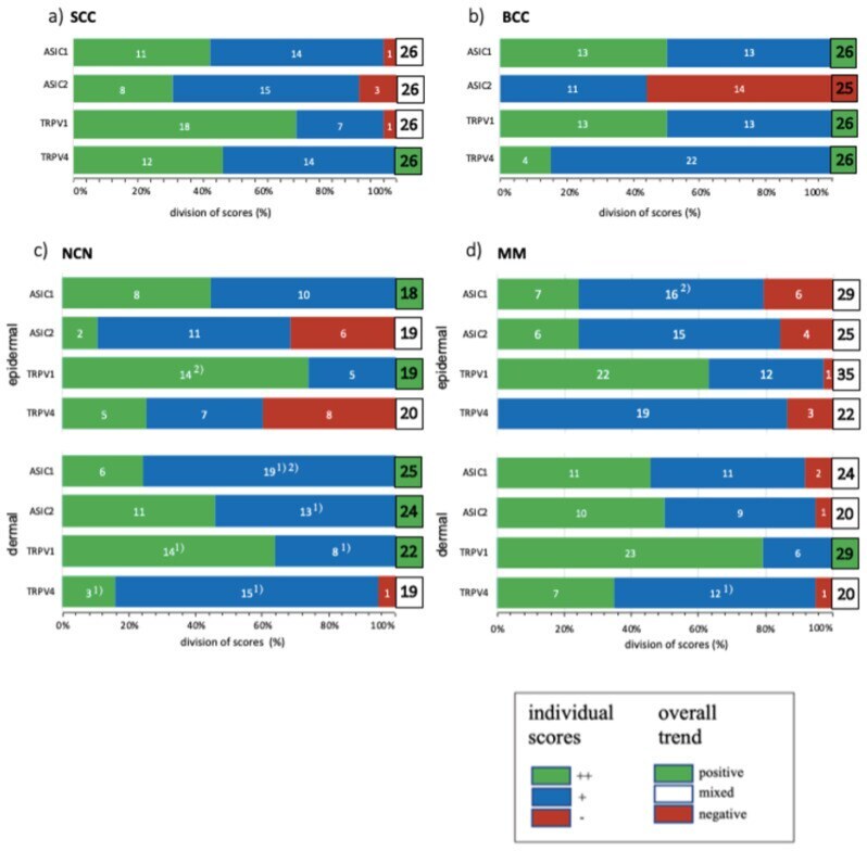
- Figure 5 Summary of standard immunohistochemical and TMA score results for ASIC1, ASIC2, TRPV1 and TRPV4 on ( a ) SCC, ( b ) BCC, ( c ) NCN and ( d ) MM. ++/green bar: strong positive staining with >80% of cells positive and/or staining intensity is high; +/blue bar: 20-80% of cells show a weak positive/partial positive reaction; -/red bar:
- Submitted by
- Invitrogen Antibodies (provider)
- Main image
- Experimental details
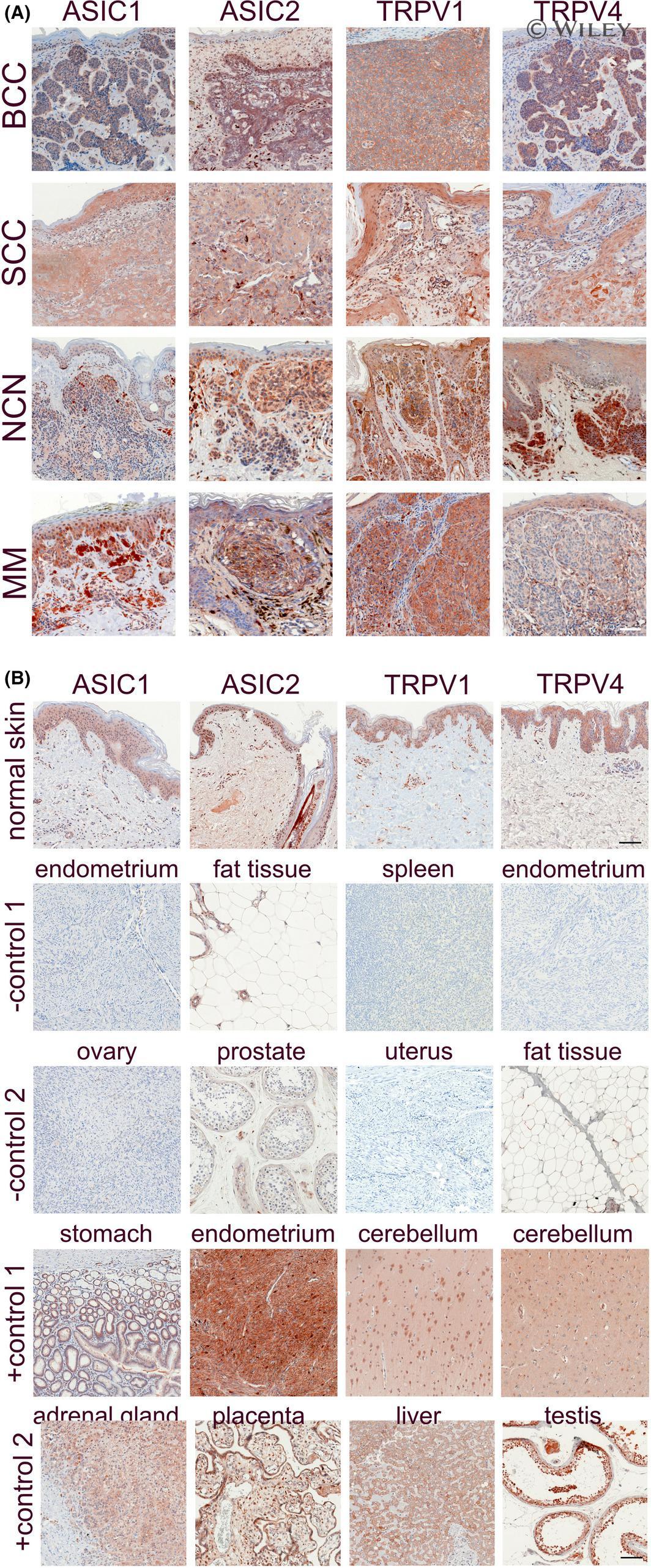
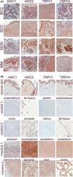
- 1 FIGURE Immunohistochemistry of ASIC1/2 and TRPV1/4: (A) Representative samples of the most common skin tumors, that is basal cell carcinoma (BCC), squamous cell carcinoma (SCC), naevus cell naevus (NCN) and melanoma (MM) were stained for ASIC1, ASIC2, TRPV1 or TRPV4, according to the protocol as described in Nassios et al [ 36 ] and using the following primary antibodies: ASIC1: PA5-26778, ASIC2: PA5-26222, TRPV1: LS-B12677 (Thermo Fisher Scientific Inc, Waltham, MA, USA), TRPV4: ab21912 (abcam, Cambridge, UK). B, Normal skin (first row) and control tissues (second to fifth row) were stained for ASIC1, ASIC2, TRPV1 or TRPV4. Negative (second and third row) and positive (fourth and fifth row) controls were selected according to expression analyses published at the human protein atlas ( https://www.proteinatlas.org/ ). Scale bars: 100 um. Compared to epidermis, BCC showed strong expression of the four pH-sensitive proteins. In contrast, SCC showed a quite similar expression of all pH-sensitive proteins in comparison to normal epidermis, markedly weaker than BCC. For NCN and MM, epidermal and dermal expression was quite different. NCN exhibit strong expression of ASIC1/2 in the more superficial portion, while the intensity decreases in dermal melanocytes. However, TRPV1/4 seems to be uniformly expressed even in deeper tissue layers. In MM, ASIC1/2 and also TRPV1 are also expressed throughout the whole tumor. In contrast, TRPV4 staining showed only weak expression in MM, which