Explore
 Validate
 Learn
Learn50-7077-41
antibody from Invitrogen Antibodies
Targeting: CCL7
FIC, MARC, MCP-3, MCP3, NC28, SCYA6, SCYA7
 Flow cytometry
Antibody data
- Antibody Data
- Antigen structure
- References [3]
- Comments [0]
- Validations
- Flow cytometry [2]
Submit
Validation data
Reference
Comment
Report error
- Product number
- 50-7077-41 - Provider product page

- Provider
- Invitrogen Antibodies
- Product name
- CCL7 (MCP-3) Monoclonal Antibody (OLGMASCE), eFluor™ 660, eBioscience™
- Antibody type
- Monoclonal
- Antigen
- Other
- Description
- Description: This OLGMASCE monoclonal antibody reacts with human CCL7. CCL7, also known as MCP-3 (Monocyte Chemotactic Protein 3), is a member of the CC- subfamily of chemokines and is most closely related to CCL2 (MCP-1) and CCL8 (MCP-2). They are secreted by a variety of cell types in response to inflammatory stimuli and play critical roles in the recruitment of leukocytes to areas of inflammation. While all three MCP proteins are potent chemoattractants of monocytes and T cells, CCL7 appears to have the broadest range of activity, as it has also been demonstrated to attract activated NK cells, eosinophils, basophils, and neutrophils. CCL7 signals via the g protein-coupled receptors CCR1 and CCR2, both of which are shared with other CC-chemokines. Applications Reported: This OLGMASCE antibody has been reported for use in intracellular staining followed by flow cytometric analysis. Applications Tested: This OLGMASCE antibody has been pre-titrated and tested by intracellular staining followed by flow cytometric analysis of stimulated normal human peripheral blood cells using the Intracellular Fixation and Permeabilization Buffer Set (Product # 88-8824-00) and protocol. This can be used at 5 µL (0.06 µg) per test. A test is defined as the amount (µg) of antibody that will stain a cell sample in a final volume of 100 µL. Cell number should be determined empirically but can range from 10^5 to 10^8 cells/test. eFluor® 660 is a replacement for Alexa Fluor® 647. eFluor® 660 emits at 659 nm and is excited with the red laser (633 nm). Please make sure that your instrument is capable of detecting this fluorochome. Excitation: 633-647 nm; Emission: 668 nm; Laser: Red Laser. Filtration: 0.2 µm post-manufacturing filtered.
- Reactivity
- Human
- Host
- Mouse
- Isotype
- IgG
- Antibody clone number
- OLGMASCE
- Vial size
- 25 Tests
- Concentration
- 5 μL/Test
- Storage
- 4°C, store in dark, DO NOT FREEZE!
Submitted references Inflammatory cytokines stimulate the chemokines CCL2/MCP-1 and CCL7/MCP-3 through NFkB and MAPK dependent pathways in rat astrocytes [corrected].
Additive roles for MCP-1 and MCP-3 in CCR2-mediated recruitment of inflammatory monocytes during Listeria monocytogenes infection.
Human monocyte chemotactic proteins-2 and -3: structural and functional comparison with MCP-1.
Thompson WL, Van Eldik LJ
Brain research 2009 Sep 1;1287:47-57
Brain research 2009 Sep 1;1287:47-57
Additive roles for MCP-1 and MCP-3 in CCR2-mediated recruitment of inflammatory monocytes during Listeria monocytogenes infection.
Jia T, Serbina NV, Brandl K, Zhong MX, Leiner IM, Charo IF, Pamer EG
Journal of immunology (Baltimore, Md. : 1950) 2008 May 15;180(10):6846-53
Journal of immunology (Baltimore, Md. : 1950) 2008 May 15;180(10):6846-53
Human monocyte chemotactic proteins-2 and -3: structural and functional comparison with MCP-1.
Proost P, Wuyts A, Van Damme J
Journal of leukocyte biology 1996 Jan;59(1):67-74
Journal of leukocyte biology 1996 Jan;59(1):67-74
No comments: Submit comment
Supportive validation
- Submitted by
- Invitrogen Antibodies (provider)
- Main image
- Experimental details
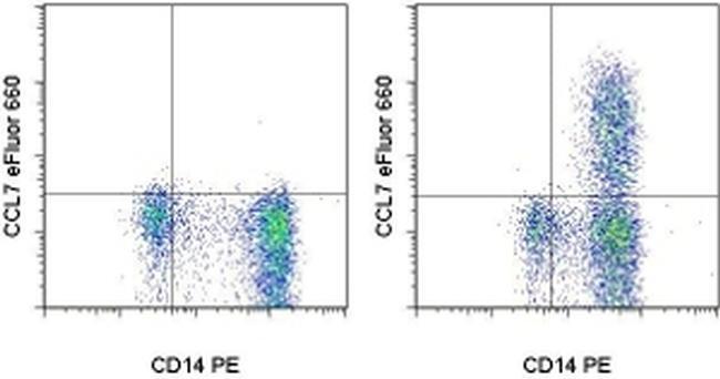
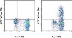
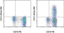
- Normal human peripheral blood cells were unstimulated (left) or stimulated 5 hours with LPS in the presence of Protein Transport Inhibitor Cocktail (Product # 00-4980-03) (right). Cells were intracellularly stained with Anti-Human CD14 PE (Product # 12-0149-42) and Anti-Human CCL7 eFluor® 660 using the Intracellular Fixation and Permeabilization Buffer Set (Product # 88-8824-00) and protocol. Cells in the monocyte gate were used for analysis.

- Submitted by
- Invitrogen Antibodies (provider)
- Main image
- Experimental details
- Normal human peripheral blood cells were unstimulated (left) or stimulated 5 hours with LPS in the presence of Protein Transport Inhibitor Cocktail (Product # 00-4980-03) (right). Cells were intracellularly stained with Anti-Human CD14 PE (Product # 12-0149-42) and Anti-Human CCL7 eFluor® 660 using the Intracellular Fixation and Permeabilization Buffer Set (Product # 88-8824-00) and protocol. Cells in the monocyte gate were used for analysis.