Explore
 Validate
 Learn
LearnMA5-29089
antibody from Invitrogen Antibodies
Targeting: CCL7
FIC, MARC, MCP-3, MCP3, NC28, SCYA6, SCYA7
 ELISA
ELISA Immunocytochemistry
Antibody data
- Antibody Data
- Antigen structure
- References [1]
- Comments [0]
- Validations
- Immunocytochemistry [1]
- Flow cytometry [2]
- Other assay [3]
Submit
Validation data
Reference
Comment
Report error
- Product number
- MA5-29089 - Provider product page

- Provider
- Invitrogen Antibodies
- Product name
- MCP-3 Monoclonal Antibody (18)
- Antibody type
- Monoclonal
- Antigen
- Recombinant full-length protein
- Description
- This product is preservative free. It is recommended to add sodium azide to avoid contamination (final concentration 0.05%-0.1%). This antibody has specificity for Human CCL7/MCP3.
- Reactivity
- Human
- Host
- Mouse
- Isotype
- IgG
- Antibody clone number
- 18
- Vial size
- 100 μL
- Concentration
- 1 mg/mL
- Storage
- Store at 4°C short term. For long term storage, store at -20°C, avoiding freeze/thaw cycles.
Submitted references LINC01094/SPI1/CCL7 Axis Promotes Macrophage Accumulation in Lung Adenocarcinoma and Tumor Cell Dissemination.
Wu Z, Bai X, Lu Z, Liu S, Jiang H
Journal of immunology research 2022;2022:6450721
Journal of immunology research 2022;2022:6450721
No comments: Submit comment
Supportive validation
- Submitted by
- Invitrogen Antibodies (provider)
- Main image
- Experimental details
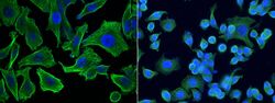
- Immunofluorescence staining of Human MCP-3 in SKBR3 cells. Cells were fixed with 4% PFA, permeabilzed with 1% Triton X-100 in PBS, blocked with 10% serum, and incubated with MCP-3 Monoclonal Antibody (18) (Product # MA5-29089, 1:60). Then cells were stained with the Alexa Fluor® 488-conjugated Goat Anti-mouse IgG secondary antibody (left panel, captured by laser confocal scanning microscope; right panel, captured by fluorescence microscope), counterstained with DAPI (blue). Positive staining was localized to cytoplasm.
Supportive validation
- Submitted by
- Invitrogen Antibodies (provider)
- Main image

- Experimental details
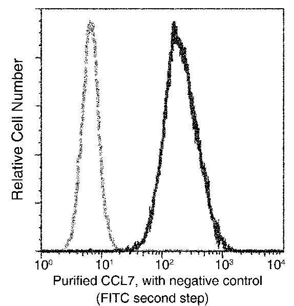
- Flow cytometric analysis of Human MCP-3 expression on THP-1 cells. The cells were treated according to manufacturer’s manual, stained with MCP-3 Monoclonal Antibody (18) (Product # MA5-29089), then a FITC-conjugated Secondary antibody. The fluorescence histograms were derived from gated events with the forward and side light-scatter characteristics of intact cells.

- Submitted by
- Invitrogen Antibodies (provider)
- Main image

- Experimental details
- Flow cytometric analysis of Human MCP-3 expression on THP-1 cells. The cells were treated according to manufacturer’s manual, stained with MCP-3 Monoclonal Antibody (18) (Product # MA5-29089), then a FITC-conjugated Secondary antibody. The fluorescence histograms were derived from gated events with the forward and side light-scatter characteristics of intact cells.
Supportive validation
- Submitted by
- Invitrogen Antibodies (provider)
- Main image
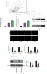
- Experimental details
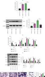
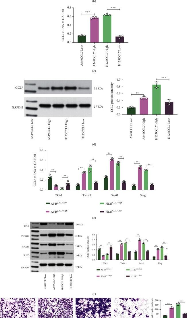
- Figure 2 CCL7 knockdown suppresses EMT and mobility of LUAD cells. mRNA (a) and protein (b) levels of CCL7 in H1299, A549, H358 (low-invasive), and H125 (high-invasive) cells detected by qPCR and WB analyses; mRNA (c) and protein (d) levels of CCL7 in A549 and H125 cells after overexpression plasmid or shRNA transfection determined by qPCR and WB analyses; mRNA (e) and protein (f) levels of epithelial marker ZO-1 and mesenchymal markers Twist1, Snai1, and Slug in A549 and H125 cells examined by qPCR and WB analyses; (g) 24-h migration rate of A549 and H125 cells analyzed by the scratch test; (h) invasiveness of A549 and H125 cells analyzed by Transwell assay. Repetition = 3. ** p < 0.01.
- Submitted by
- Invitrogen Antibodies (provider)
- Main image
- Experimental details
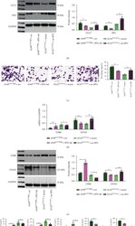
- Overexpression of SPI1 in CCL7-low cells promotes macrophage migration and M2 polarization. mRNA (a) and protein (b) levels of SPI1 and CCL7 in H125 CCL7 Low and A549 CCL7 High cells after SPI1 overexpression plasmid or shRNA transfection detected by qPCR and WB analyses; (c) number of migrated THP-1 cells examined by crystal violet staining; mRNA (d) and protein (e) levels of CD86 and CD206 in cocultured THP-1 cells examined by qPCR and WB analyses; (f) expression of M1 cytokines (IL-6, IL-1 beta , and TNF- alpha ) and M2 cytokines (IL-10 and TGF- beta ) in the culture medium determined by ELISA kits. Repetition = 3. Data are presented as the mean +- SD. ** p < 0.01.
- Submitted by
- Invitrogen Antibodies (provider)
- Main image
- Experimental details
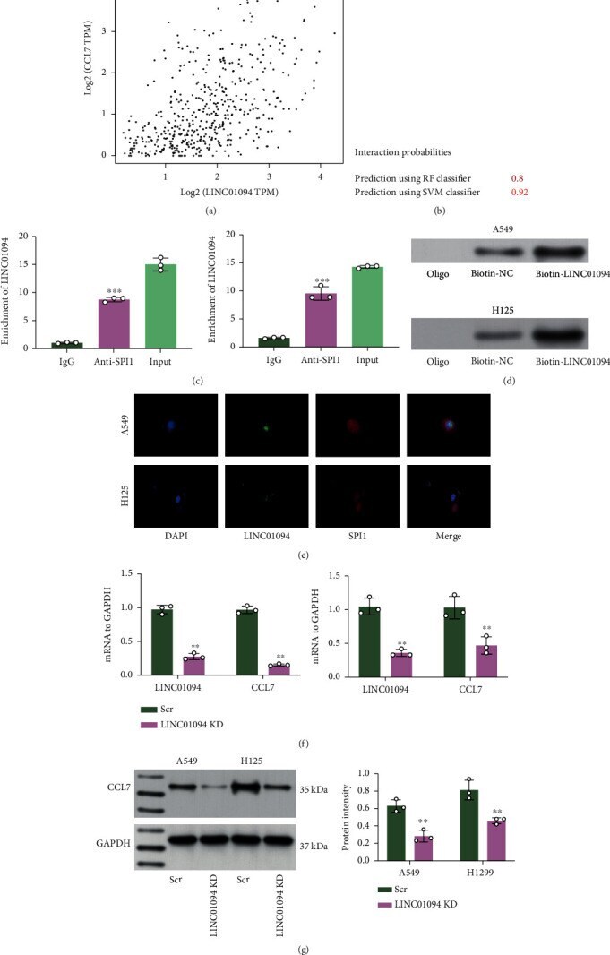
- Figure 7 LINC01094 binds to SPI1 to regulate CCL7 expression. (a) Correlation of LINC01094 with CCL7 predicted in the TCGA-LUAD database; (b) binding relationship between LINC01094 and SPI1 validated in TCGA-LUAD database; binding between LINC01094 and SPI1 examined by RIP (c) and biotin-LINC01094-based RNA pull down (d) assays; (e) subcellular localization of LINC01094 and SPI1 in H125 and A549 cells examined by FISH assay; mRNA (f) and protein (g) levels of CCL7 in A549 and H125 cells after LINC01094 knockdown examined by qPCR and WB analyses. Repetition = 3. Data are presented as the mean +- SD. ** p < 0.01.