 Explore
 Validate
 Learn
Learn Western blot
Antibody data
- Antibody Data
- Antigen structure
- References [1]
- Comments [0]
- Validations
- Western blot [6]
- Immunocytochemistry [2]
- Immunohistochemistry [3]
- Other assay [1]
Submit
Validation data
Reference
Comment
Report error
- Product number
- PA5-21290 - Provider product page

- Provider
- Invitrogen Antibodies
- Product name
- NQO1 Polyclonal Antibody
- Antibody type
- Polyclonal
- Antigen
- Synthetic peptide
- Description
- Recommended positive controls: A431, HeLa, HepG2, mouse heart, rat heart. Predicted reactivity: Rhesus Monkey (100%). Store product as a concentrated solution. Centrifuge briefly prior to opening the vial.
- Reactivity
- Human, Mouse, Rat
- Host
- Rabbit
- Isotype
- IgG
- Vial size
- 100 µL
- Concentration
- 0.61 mg/mL
- Storage
- Store at 4°C short term. For long term storage, store at -20°C, avoiding freeze/thaw cycles.
Submitted references Cancer Chemopreventive Potential and Chemical Profiling of Euphorbia abyssinica Endowed with Docking Studies.
Ahmed SR, Hamed AR, Ali MI, Sedeek MS, Abelyan N, Al-Sanea MM
ACS omega 2022 Feb 1;7(4):3596-3604
ACS omega 2022 Feb 1;7(4):3596-3604
No comments: Submit comment
Supportive validation
- Submitted by
- Invitrogen Antibodies (provider)
- Main image
- Experimental details
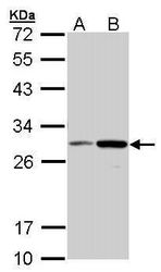
- Western blot analysis of NQO1 using 30 µg of A) MOLT4 and B) Raji lysate. Samples were loaded onto a 12% SDS-PAGE gel and probed with a NQO1 polyclonal antibody (Product # PA5-21290) at a dilution of 1:1000.

- Submitted by
- Invitrogen Antibodies (provider)
- Main image
- Experimental details
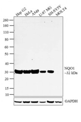
- Western blot analysis was performed on whole cell extracts (30 µg lysate) of Hep G2 (Lane 1), HeLa (Lane 2), A549 (Lane 3), U-87 MG (Lane 4), SH-SY5Y (Lane 5) and MOLT4 (Lane 6). The blot was probed with Anti-NQO1 Polyclonal Antibody (Product # PA5-21290, 1:2000 dilution) and detected by chemiluminescence using Goat anti Rabbit IgG (H+L) Superclonal™ Secondary Antibody, HRP conjugate (Product # A27036, 0.25 µg/ml, 1:4000 dilution). A 32 kDa band corresponding to NQO1 was detected across the cell lines tested except SH-SY 5Y and MOLT4 which showed little or no expression respectively as reported in the literature.

- Submitted by
- Invitrogen Antibodies (provider)
- Main image
- Experimental details
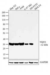
- Western blot analysis was performed on whole cell extracts (30 µg lysate) of Hep G2 (Lane 1), HeLa (Lane 2), A549 (Lane 3), U-87 MG (Lane 4), SH-SY5Y (Lane 5) and MOLT4 (Lane 6). The blot was probed with Anti-NQO1 Polyclonal Antibody (Product # PA5-21290, 1:2000 dilution) and detected by chemiluminescence using Goat anti Rabbit IgG (H+L) Superclonal™ Secondary Antibody, HRP conjugate (Product # A27036, 0.25 µg/ml, 1:4000 dilution). A 32 kDa band corresponding to NQO1 was detected across the cell lines tested except SH-SY 5Y and MOLT4 which showed little or no expression respectively as reported in the literature.

- Submitted by
- Invitrogen Antibodies (provider)
- Main image
- Experimental details
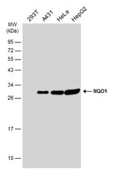
- Western Blot analysis of NQO1 was performed by separating 30 µg of various whole cell extracts by 12% SDS-PAGE. Proteins were transferred to a membrane and probed with a NQO1 Polyclonal Antibody (Product # PA5-21290) at a dilution of 1:1000 and a HRP-conjugated anti-rabbit IgG secondary antibody.

- Submitted by
- Invitrogen Antibodies (provider)
- Main image
- Experimental details
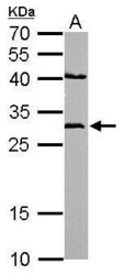
- NQO1 Polyclonal Antibody detects NQO1 protein by western blot analysis. A. 50 µg mouse heart lysate/extract.12% SDS-PAGE. NQO1 Polyclonal Antibody (Product # PA5-21290) dilution: 1:500. The HRP-conjugated anti-rabbit IgG antibody was used to detect the primary antibody.

- Submitted by
- Invitrogen Antibodies (provider)
- Main image
- Experimental details
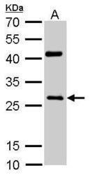
- NQO1 Polyclonal Antibody detects NQO1 protein by western blot analysis. A. 50 µg rat heart lysate/extract.12% SDS-PAGE. NQO1 Polyclonal Antibody (Product # PA5-21290) dilution: 1:500. The HRP-conjugated anti-rabbit IgG antibody was used to detect the primary antibody.

Supportive validation
- Submitted by
- Invitrogen Antibodies (provider)
- Main image

- Experimental details
- Immunofluorescent analysis of NQO1 in paraformaldehyde-fixed HeLa cells using a NQO1 polyclonal antibody (Product # PA5-21290) at a 1:200 dilution.

- Submitted by
- Invitrogen Antibodies (provider)
- Main image

- Experimental details
- Immunofluorescence analysis of paraformaldehyde-fixed HeLa, using NQO1 antibody (Product # PA5-21290) at 1:200 dilution.

Supportive validation
- Submitted by
- Invitrogen Antibodies (provider)
- Main image
- Experimental details
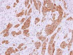
- Immunohistochemistry (Paraffin) analysis of NQO1 was performed in paraffin-embedded human rectal carcinoma tissue using NQO1 Polyclonal Antibody (Product # PA5-21290) at a dilution of 1:500.

- Submitted by
- Invitrogen Antibodies (provider)
- Main image

- Experimental details
- Immunohistochemistry (Paraffin) analysis of NQO1 was performed in paraffin-embedded mouse kidney tissue using NQO1 Polyclonal Antibody (Product # PA5-21290) at a dilution of 1:500. Antigen Retrieval: Citrate buffer, pH 6.0, 15 min.

- Submitted by
- Invitrogen Antibodies (provider)
- Main image

- Experimental details
- NQO1 Polyclonal Antibody detects NQO1 protein at cytoplasm by immunohistochemical analysis. Sample: Paraffin-embedded mouse kidney. NQO1 stained by NQO1 Polyclonal Antibody (Product # PA5-21290) diluted at 1:500. Antigen Retrieval: Citrate buffer, pH 6.0, 15 min.

Supportive validation
- Submitted by
- Invitrogen Antibodies (provider)
- Main image

- Experimental details
- Figure 4 Western blotting showing concentration-dependent upregulation of the chemopreventive marker NQO1 protein expressions by the Euphorbia abyssinica fruit extract in a Hepa1c1c7 cell line.
