Explore
 Validate
 Learn
LearnMA1-16672
antibody from Invitrogen Antibodies
Targeting: NQO1
DHQU, DIA4, DTD, NMOR1, QR1
 Western blot
 Immunocytochemistry
 Immunoprecipitation Immunohistochemistry Flow cytometry Other assay
Antibody data
- Antibody Data
- Antigen structure
- References [2]
- Comments [0]
- Validations
- Immunocytochemistry [2]
- Immunohistochemistry [2]
- Flow cytometry [8]
- Other assay [2]
Submit
Validation data
Reference
Comment
Report error
- Product number
- MA1-16672 - Provider product page

- Provider
- Invitrogen Antibodies
- Product name
- NQO1 Monoclonal Antibody (A180)
- Antibody type
- Monoclonal
- Antigen
- Recombinant full-length protein
- Description
- This antibody reacts weakly with mouse tissue.
- Reactivity
- Human, Mouse, Rat, Canine
- Host
- Mouse
- Isotype
- IgG
- Antibody clone number
- A180
- Vial size
- 100 μL
- Concentration
- 1 mg/mL
- Storage
- Store at 4°C short term. For long term storage, store at -20°C, avoiding freeze/thaw cycles.
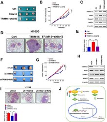
Submitted references E3 ligase TRIM15 facilitates non-small cell lung cancer progression through mediating Keap1-Nrf2 signaling pathway.
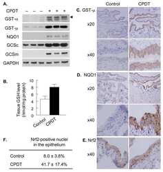
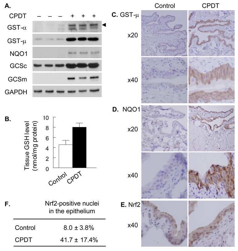
5,6-Dihydrocyclopenta[c][1,2]-dithiole-3(4H)-thione is a promising cancer chemopreventive agent in the urinary bladder.
Liang M, Wang L, Sun Z, Chen X, Wang H, Qin L, Zhao W, Geng B
Cell communication and signaling : CCS 2022 May 9;20(1):62
Cell communication and signaling : CCS 2022 May 9;20(1):62
5,6-Dihydrocyclopenta[c][1,2]-dithiole-3(4H)-thione is a promising cancer chemopreventive agent in the urinary bladder.
Paonessa JD, Munday CM, Mhawech-Fauceglia P, Munday R, Zhang Y
Chemico-biological interactions 2009 Jun 15;180(1):119-26
Chemico-biological interactions 2009 Jun 15;180(1):119-26
No comments: Submit comment
Supportive validation
- Submitted by
- Invitrogen Antibodies (provider)
- Main image
- Experimental details
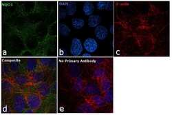
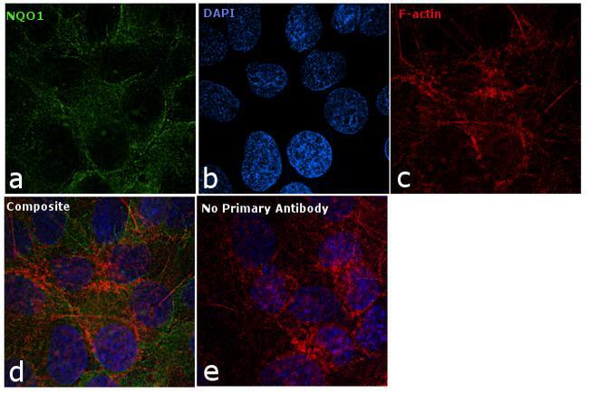
- Immunofluorescence analysis of NQO1 was performed using 70% confluent log phase Hep G2 cells. The cells were fixed with 4% paraformaldehyde for 10 minutes, permeabilized with 0.1% Triton™ X-100 for 10 minutes, and blocked with 1% BSA for 1 hour at room temperature. The cells were labeled with NQO1 Mouse Monoclonal Antibody (A180) (Product # MA1-16672) at 5 µg/mL in 0.1% BSA and incubated overnight at 4 degree and then labeled with Goat anti-Mouse IgG (H+L) Superclonal™ Secondary Antibody, Alexa Fluor® 488 conjugate (Product # A28175) at a dilution of 1:2000 for 45 minutes at room temperature (Panel a: green). Nuclei (Panel b: blue) were stained with SlowFade® Gold Antifade Mountant with DAPI (Product # S36938). F-actin (Panel c: red) was stained with Rhodamine Phalloidin (Product # R415, 1:300). Panel d represents the merged image showing cytoplasmic localization. Panel e represents control cells with no primary antibody to assess background. The images were captured at 60X magnification.

- Submitted by
- Invitrogen Antibodies (provider)
- Main image

- Experimental details
- Immunofluorescence analysis of NQO1 was performed using 70% confluent log phase Hep G2 cells. The cells were fixed with 4% paraformaldehyde for 10 minutes, permeabilized with 0.1% Triton™ X-100 for 10 minutes, and blocked with 1% BSA for 1 hour at room temperature. The cells were labeled with NQO1 Mouse Monoclonal Antibody (A180) (Product # MA1-16672) at 5 µg/mL in 0.1% BSA and incubated overnight at 4 degree and then labeled with Goat anti-Mouse IgG (H+L) Superclonal™ Secondary Antibody, Alexa Fluor® 488 conjugate (Product # A28175) at a dilution of 1:2000 for 45 minutes at room temperature (Panel a: green). Nuclei (Panel b: blue) were stained with SlowFade® Gold Antifade Mountant with DAPI (Product # S36938). F-actin (Panel c: red) was stained with Rhodamine Phalloidin (Product # R415, 1:300). Panel d represents the merged image showing cytoplasmic localization. Panel e represents control cells with no primary antibody to assess background. The images were captured at 60X magnification.
Supportive validation
- Submitted by
- Invitrogen Antibodies (provider)
- Main image
- Experimental details
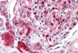
- Immunohistochemical analysis of NQO1 in formalin fixed paraffin embedded human kidney tissue. Samples were incubated in NQO1 monoclonal antibody (Product # MA1-16672).

- Submitted by
- Invitrogen Antibodies (provider)
- Main image

- Experimental details
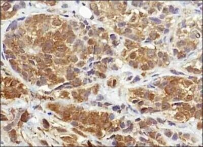
- Immunohistochemical analysis of NQO1 in human breast cancer. Samples were incubated in NQO1 monoclonal antibody (Product # MA1-16672) followed by DAB with hematoxylin counterstain.
Supportive validation
- Submitted by
- Invitrogen Antibodies (provider)
- Main image

- Experimental details
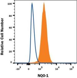
- Flow cytometry of NQO1 in Human A549 cell line. Samples were incubated in NQO1 monoclonal antibody (Product # MA1-16672) or Mouse IgG1 isotype control followed by APC-conjugated Anti-Mouse IgG Secondary Antibody. To facilitate intracellular staining, cells were fixed with FlowX FoxP3 Fixation & Permeabilization Buffer Kit.

- Submitted by
- Invitrogen Antibodies (provider)
- Main image
- Experimental details
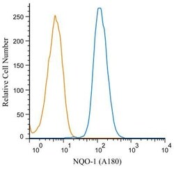
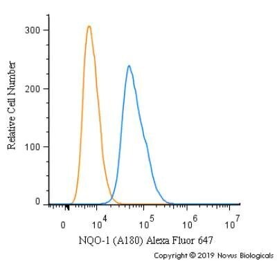
- Flow cytometry of NQO1 in U87MG cells. Samples were incubated in NQO1 monoclonal antibody (Product # MA1-16672) along with a matched isotype control using a dilution of 1 µg/mL for 30 minutes at room temperature followed by mouse F(ab)2 IgG (H+L) APC-conjugated secondary antibody. Antibody (blue) along with a matched isotype control (orange). Cells were fixed with 4% PFA and permeabilized with 0.1% saponin.

- Submitted by
- Invitrogen Antibodies (provider)
- Main image
- Experimental details
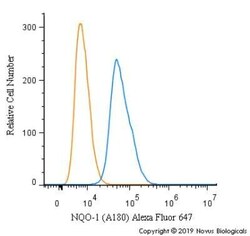
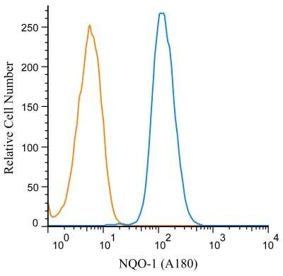
- Flow cytometry of NQO1 in U87 cells. Samples were incubated in NQO1 monoclonal antibody (Product # MA1-16672) using a dilution of 2.5 µg/mL for 30 minutes at room temperature. Antibody (blue) and a matched isotype control (orange). Cells were fixed with 4% PFA and then permeabilized with 0.1% saponin. Both antibodies were conjugated to Alexa Fluor 647.

- Submitted by
- Invitrogen Antibodies (provider)
- Main image

- Experimental details
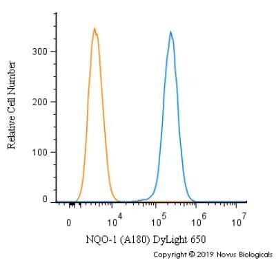
- Flow cytometry of NQO1 in U-87 cells. Samples were incubated in NQO1 monoclonal antibody (Product # MA1-16672) using a dilution of 2.5 µg/mL for 30 minutes at room temperature. Antibody (blue) and a matched isotype control (orange). Cells were fixed with 4% PFA and then permeabilized with 0.1% saponin. Both antibodies were conjugated to DyLight 650.

- Submitted by
- Invitrogen Antibodies (provider)
- Main image
- Experimental details
- Flow cytometry of NQO1 in Human A549 cell line. Samples were incubated in NQO1 monoclonal antibody (Product # MA1-16672) or Mouse IgG1 isotype control followed by APC-conjugated Anti-Mouse IgG Secondary Antibody. To facilitate intracellular staining, cells were fixed with FlowX FoxP3 Fixation & Permeabilization Buffer Kit.

- Submitted by
- Invitrogen Antibodies (provider)
- Main image

- Experimental details
- Flow cytometry of NQO1 in U87MG cells. Samples were incubated in NQO1 monoclonal antibody (Product # MA1-16672) along with a matched isotype control using a dilution of 1 µg/mL for 30 minutes at room temperature followed by mouse F(ab)2 IgG (H+L) APC-conjugated secondary antibody. Antibody (blue) along with a matched isotype control (orange). Cells were fixed with 4% PFA and permeabilized with 0.1% saponin.
- Submitted by
- Invitrogen Antibodies (provider)
- Main image

- Experimental details
- Flow cytometry of NQO1 in U87 cells. Samples were incubated in NQO1 monoclonal antibody (Product # MA1-16672) using a dilution of 2.5 µg/mL for 30 minutes at room temperature. Antibody (blue) and a matched isotype control (orange). Cells were fixed with 4% PFA and then permeabilized with 0.1% saponin. Both antibodies were conjugated to Alexa Fluor 647.
- Submitted by
- Invitrogen Antibodies (provider)
- Main image
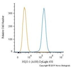
- Experimental details
- Flow cytometry of NQO1 in U-87 cells. Samples were incubated in NQO1 monoclonal antibody (Product # MA1-16672) using a dilution of 2.5 µg/mL for 30 minutes at room temperature. Antibody (blue) and a matched isotype control (orange). Cells were fixed with 4% PFA and then permeabilized with 0.1% saponin. Both antibodies were conjugated to DyLight 650.
Supportive validation
- Submitted by
- Invitrogen Antibodies (provider)
- Main image
- Experimental details
- NULL
- Submitted by
- Invitrogen Antibodies (provider)
- Main image
- Experimental details
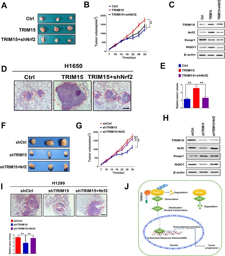
- Fig. 7 TRIM15 mediated increase in Nrf2 regulates growth and invasion in vivo. A Nude mice were randomized into three groups and subcutaneously injected with H1650 cells that had been transfected with control (empty vector), TRIM15, or TRIM15 + shNrf2 plasmids. Tumors formed in nude mice were collected 30 days after grafting, and the tumor weight were measured. B Measurement of tumor volume in experimental groups over time. C Western blotting analysis was performed to evaluate the levels of TRIM15, Nrf2, Keap1, and NQO1 in harvested tumors. D , E Up-regulation of TRIM15 significantly promoted lung metastasis in H1650 xenograft nude mice models, whereas the suppression of Nrf2 prevented the tumor metastasis of TRIM15 overexpressing cells. Representative pictures of the lung metastases in nude mice by H&E staining. Quantification of lung metastases in all groups. Scale bar: 200 mum. F , G A representative image of tumor growth in nude mice subcutaneously inoculated with H1299 cells tranfected with shCtrl, shTRIM15 or shTRIM15 + Nrf2 plasmids. Tumor volumes were measured on the indicated days. H Western blotting analysis was performed to evaluate the levels of TRIM15, Nrf2, Keap1, and NQO1 in xenograft tumors. I Representative pictures of the lung metastases in nude mice by H&E staining. Quantification of lung metastases in all groups. Scale bar: 200 mum. J TRIM15 was significantly upregulated in NSCLC and that increased TRIM15 was associated with poor survival. TRIM15 promoted